All posts by The Trauma Pro

Advanced Needle Thoracostomy

I’ve recently written about the merits of needle vs finger thoracostomy. One of the arguments against needle thoracostomy is that it may not reach into the chest cavity in obese patients. As I mentioned yesterday, use the right needle!

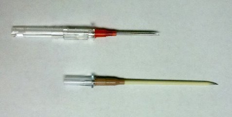
Obviously, the one on top isn’t going to get you very far. The bottom one (10 gauge 3 inch) should get into most pleural spaces.

But what if you don’t have the right needle? Or what if the patient is massively obese and the longer needle won’t even reach? Pushing harder may seem logical, but it doesn’t work. You might be able to get the needle to reach to the pleural space, but the catheter won’t stay in it.

Here’s the trick. First, make the angiocatheter longer by hooking it up to a small (5 or 10cc) syringe. Now prep the chest over your location of choice (2nd intercostal space, mid-clavicular line or 5th intercostal space, anterior axillary line) and make a skin incision slightly larger than the diameter of the syringe. Now place the syringe and attached needle into the chest via your incision. It is guaranteed to reach the pleura, because you can now get the hub of the catheter down to the level of the ribs. Just don’t forget to pull out the catheter once you’ve placed the chest tube!

Related posts:

Why I Don’t Like Finger Thoracostomy

I continue to see interest in using finger thoracostomy in place of needling the chest for the management of real or presumed tension pneumothorax. As noted in the title, I don’t really care for this procedure. I know that there’s a lot of opinion on this topic out there, especially in blogs. My colleague, Scott Weingart (EMCrit) has a very nice podcast on the topic (link below). But the actual scientific literature supporting or condemning its use is sparse.

The procedure consists of doing a limited prep of the chest in the same location for regular chest tube placement, incision, rapid puncture of the parietal pleura, followed by placement of a finger into the pleural space to release tension. Sounds well and good! So what’s my beef? The arguments for it emphasize speed, certainty, and reversibility.

Let’s talk about speed first. This procedure is supposed to be fast. An incision, a few quick sweeps with a clamp, and voila! Finger inserted. And it can be this fast. But in reality, especially in training centers, people who don’t insert chest tubes very often take too long (1-2 minutes).

The next argument is certainty. There are a number of papers showing that needle thoracostomies often miss the mark, especially when using standard through the catheter needles. This is more likely to occur when the needle is inserted in the standard location (2nd intercostal space, midclavicular line) and in obese patients. My response is, use a longer needle!

The angio-catheter on top is a standard 14Ga 1.25 inch model, and won’t get you anywhere. It’s only good for thin people, and will kink as soon as the needle is withdrawn. The bottom model is 10Ga 3 inch, and is effective in everyone save the very morbidly obese. It’s thick and will not kink until it gets good and warm.

The final issue is reversibility. The argument goes, stick a needle in the lung and you’ll get a pneumothorax, but stick a finger in the chest and no harm done. I don’t completely buy this. Puncturing the lung does not a guarantee a pneumothorax. But it will require a subsequent chest xray to see if one develops. Finger thoracostomy doesn’t guarantee that a pneumothorax won’t occur. It also requires a chest xray later to check.

Bottom line: As you can tell, I’m not a big fan of finger thoracostomy, mainly due to speed (or lack thereof). Just stock some big fat needle catheters in your trauma bay and be done with it. But if you really, really want to use the finger technique, make sure that the person doing it is very experienced. This is not a learner’s procedure. It should take no more than 15 seconds, or the wrong person is doing it.

Related posts:

The Passing Of The Rectal Exam In Trauma

It has long been standard operating procedure to perform a digital rectal exam in all major trauma patients. The belief has always been that valuable information about blood in the GI tract, the status of the urethra, and the neuro exam (rectal tone) could be gleaned from the exam.

Unfortunately, the exam also serves to antagonize or even further traumatize some patients, especially those who may be intoxicated to some degree. On a number of occasions I have seen calm patients become so agitated by the rectal that they required intubation for control.

So is it really necessary? A study in 2001 conducted over a 6 month period (1) showed that the rectal exam influenced management in only 1.2% of cases. The authors felt that there was some utility in 3 special cases:

  • Spinal cord injury – looking for sacral sparing
  • Pelvic fracture – looking for bone shards protruding into the rectum
  • Penetrating abdominal trauma – looking for gross blood

A more recent 2005 study (2) was also critical of the rectal exam and found that using “other clinical indicators” (physical exam and other diagnostic study information) was at least equivalent, changing management only 4% of the time. They concurred with the first two indications above as well.

The Bottom Line: For most major trauma patients, the rectal exam is not worth the patient aggravation it causes. I still recommend it for the 3 special cases listed above, however, as there are no equivalent exams for these potentially serious patient problems. And remember, DON’T do it while the patient is in the logroll position. No patient likes a rectal exam, so they’ll do their best to defeat your attempt at spine precautions if you have them on their side. Supine, frog-leg only.

References:
1. Porter, Urcic. Am Surg. 2001 May;67(5):438-41.
2. Esposito et al. J Trauma. 2005 Dec;59(6):1314-9.

Post-Embolization Syndrome?

Important! If your search for a trauma topic brought you here…

The Trauma Professional’s Blog has moved to this new site, but old Google or Bing searches may link you to this page.

Please retype your search in the box in the upper left corner of any page (or touch the magnifying glass above on a mobile device) to find what you are looking for.

Go to theTrauma Professional's Blog page

And hopefully, all the search engines will finish indexing the new site soon! Explore the site and enjoy! Michael

———————————————————————————-

I’ve seen a number of trauma patients who have developed pain and elevated WBC after embolization of solid organs for trauma. For kidneys and main splenic artery embolization, it’s fairly common in my experience. Turns out, this phenomenon was described in 2007-2008 in patients undergoing embolization of hepatic tumors and uterine fibroids. It was termed post-embolization syndrome, and consists of pain, fever, nausea and ileus.

An article was just published in the Journal of Trauma describing this syndrome in children after splenic embolization for blunt trauma. The authors looked at their own trauma registry over a 12 year period. Yes, it took that long to find 448 children with blunt splenic injury. Of those, only 11 underwent arterial embolization (sigh of relief).

The average age was about 13 and ISS was 16 in both groups. Kids who underwent embolization were more likely to spend some time in the ICU, had a longer hospital stay (8 vs 5 days(!)), and took longer to resume their diet (5 vs 2 days). These differences occurred despite the fact that most of the embolized children had isolated splenic injuries. Additionally, the embolized children were more likely to receive blood (3 units vs none) and plasma.

My first question about this paper is, why? Broken spleens in children do not act like broken spleens in adults. The vast majority of the cases of contrast extravasation in children stops on its own without intervention. So why did we even have to find out that post-embolization syndrome occurs in children? They shouldn’t be going through this procedure anyway! Fortunately, a deeper read of the paper provides the answer. The indication for angio was splenic pseudoaneurysm in 2, and ongoing hemorrhage in the other 9. In the case of these latter 9, it did keep the children from having their spleens operated on.

Bottom line: In general, don’t send kids for splenic angiography (99.3% of kids in this study did not have it). Ongoing hemorrhage (prior to hypotension, which is an absolute indication for OR) is probably the only indication I can think of. Pseudoaneurysm and extravasation of contrast are not indications like they are in adults. But if you do have to send them, just be aware that they may develop pain, fever and ileus that will keep them in the hospital and/or ICU for a few extra days.

Reference: Transarterial embolization in children with blunt splenic injury results in postembolization syndrome: A matched case-control study. J Trauma 73(6):1558-1563, 2012.
Practical Tip: Evaluation of Hematuria in Blunt Trauma

Bloody urine is a relatively uncommon finding in blunt trauma patients. Hematuria ranges from microscopic to gross. Microscopic means blood that can only be seen with a microscope, and gross means visible to the naked eye. In trauma, we only care about gross hematuria, which ranges from the faintest of pink to the deepest red.

In the picture above gross hematuria is present in all tubes but the far right one. Those four will need further evaluation.

In trauma, gross hematuria is a result of an injury to kidney, ureter or bladder. Blunt injury to the ureter is so rare it’s reportable, so you can pretty much forget that one unless the mechanism is extreme. So you really just need to focus on kidney and bladder.

Any victim of blunt trauma that presents with visible hematuria needs to be evaluated by CT of the abdomen and pelvis with an added CT cystogram. Standard CT technique is done without a urinary catheter, or with the catheter clamped. This is not acceptable for hematuria evaluation, as only 50% of bladder injuries show up with this technique.

CT cystogram is an add-on to the standard CT, and consists of the administration of contrast into the bladder which is then kept under pressure while the scan is performed. Delayed slices through the pelvis after the bladder is depressurized and emptied is routine. Nearly 100% of bladder injuries are detected using this technique.

If the CT shows a renal laceration or hematoma, the patient should be admitted and managed according to your solid organ injury protocol. Kidney injuries fare better that livers and spleens, and only rarely require surgery. If no kidney or bladder injury is seen, the default diagnosis of a renal contusion is the culprit. No treatment is needed, and the patient can be discharged if no other injuries are present. The blood will clear over a few days, but may disappear and reappear a few times in the process. Be sure to warn the patient that this may occur, or you may receive some surprise phone calls. The patient can followup with their primary care physician in a week or two.

The majority of these injuries do not require urologic consultation. Complex injuries with extravasation of urine out of the kidney, or injuries to the collecting system should be referred to a urologist, however.