All posts by The Trauma Pro

Retained Hemothorax Part 3: VATS

I’ve written about the use of lytics to treat retained hemothorax over the past few days. Although it sounds like a good idea, we just don’t know that it works very well. And they certainly don’t work fast. Lengths of stay were on the order of two weeks in both studies reviewed.

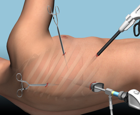
The alternative is video assisted thoracoscopic surgery (VATS). So let’s take a look at what we know about it. This procedure is basically laparoscopy of the chest. A camera is inserted, and other ports are added to allow insertion of instruments to suck, peel, and scrape out the hemothorax.

A prospective, multi-center study was performed over a 2 year period starting in 2009. Twenty centers participated, contributing data on 328 patients with retained hemothorax. This was defined as CT confirmation of retained blood and clot after chest tube placement, with evidence of pleural thickening.

Here are the factoids:

  • 41% of patients had antibiotics given for chest tube placement (this is interesting given the lack of consensus regarding their effectiveness!)
  • A third of patients were initially managed with observation, and most of them (82%) did not need any further procedures (83 of 101 patients)
  • Observation was more successful in patients who were older, had smaller hemothoraces (<300cc), smaller chest tubes (!!, <34 Fr), blunt trauma, and peri-procedure antibiotics (?)
  • An additional chest tube was inserted in 19% of patients, image guided drain placement in 5%, and lytics in 5%. Half to two-thirds of these patients required additional management.
  • VATS was used in 34% of patients. One third of them required additional management including another chest tube, another VATS, or even thoracotomy.
  • Thoracotomy was most likely required if there was a diaphragm injury or large hemothorax (<900cc)
  • Empyema and pneumonia were common (27% and 20%, respectively)

Bottom line: There’s a lot of data in this paper. Most notably, many patients resolve their hemothorax without any additional management. But if they don’t, additional tubes, guided drain placement, and lytics work only a third of the time and contribute to additional time in the hospital. Even VATS and thoracotomy require additional maneuvers 20-30% of the time. And infectious complications are common. This is a tough problem!

Tomorrow, I’ll try to roll it all together and suggest an algorithm to try to optimize both outcomes and cost.

Posts in this series:

Reference: Management of post-traumatic retained hemothorax: A prospective, observational, multicenter AAST study. 72(1):11-24, 2012.

Retained Hemothorax Part 2: Lytics (again)

Yesterday, I reviewed a small case report that was published a couple of years ago on lytics for treatment of retained hemothorax. But surely, there must be something better, right?

After digging around, I did find a paper from 2007 that prospectively looked at protocolized management of retained hemothorax, and its aftermath. It was carried out at a busy Level I trauma center over a 16 month period.

All patients with a hemothorax treated with chest tube received daily chest x-rays. Those with significant opacification on day 3 underwent CT scan of the chest. If more than 300 cc of retained blood was present, the patient received streptokinase or urokinase (surgeon preference and drug availability) daily, and rolled around in bed for 4 hours to attempt to distribute it. The chest tube was then unclamped and allowed to drain. This was repeated for 3 days, and if there was still opacification, a repeat CT was obtained. If the volume was still greater than 300 cc, the cycle was repeated for the next 3 days. If the opacification cleared at any point, or the repeat CT showed less than 300 cc, the protocol was stopped and the chest tube removed. If the chest was still opacified after 6 days, VATS was offered.

Here are the factoids:

  • A total of 203 patients with hemothorax were admitted during the study period and 25 (12%) developed a retained hemothorax
  • While a few had treatment start within 4 days, the majority did not receive lytics until day 9 (range 3  –30 days!)
  • The average length of time in hospital after start of lytics was 7 days, leading to a total length of stay of 18 days
  • 92% of patients had “effective” evacuation of their retained hemothorax, although 1 had VATS anyway which found only 100 cc of fluid
  • 16 patients had “complete” evacuation, and 5 had “partial” evacuation
  • There were no hemorrhagic complications, but one third of patients reported significant pain with drug administration

Bottom line: Sounds good, right? The drug seems reasonably effective, although lengths of stay are relatively long. However, streptokinase and urokinase are no longer available in the US, having been replaced with tissue plasminogen activator (tPA). This paper does a cost analysis of lytics vs VATS and found that the former treatment cost about $15000 (drug + hospital stay) vs $34000 for VATS. However, a big part of this was that the drug only cost about $75 per dose. tPA is much more expensive.

So once again, small series, longer lengths of stay, but at least nicely done. Unfortunately, the drug choice is no longer available so use of tPA tilts the balance away from lytics. Tomorrow, I’ll explore the results of using VATS for this condition.

Posts in this series:

Reference: Intrapleural Thrombolysis for the Management of Undrained Traumatic Hemothorax: A Prospective Observational Study. J Trauma 62(5):1175-1179, 2007.

Retained Hemothorax Part 1: Lytics

Hemothorax is a common complication of chest trauma, occurring in about one third of cases. It is commonly treated with a chest tube, which usually takes care of the problem. But in a few cases some blood remains, which can result in an entrapped lung or empyema.

There are several management options. Historically, these patients underwent thoracotomy to peel out the fibrinous collection stuck to lung and chest wall. This has given way to the more humane VATS procedure (video assisted thoracoscopic surgery) which accomplishes the same thing using a scope. In some cases, another tube can be inserted, sometimes under CT guidance, to try to drain the blood.

So what about lytics? It’s fibrin, right? So why not just dissolve it with tissue plasminogen activator (tPA)? There have been very few studies published over the years. The most recent was in 2014. I’ll review it today, and another tomorrow. Finally, I’ll give you my thoughts on the best way to deal with retained hemothorax.

Here are the factoids:

  • This was a single center, retrospective review of data from 1.5 years beginning in 2009
  • A total of seven patients were identified, and most had hemothorax due to rib fractures. Three presented immediately after their injury, 4 were delayed.
  • Median time from injury to chest tube placement was 11 days
  • Median time the chest tube was in place was 13 days, with an average hospital stay of 14 days
  • Patients received 1 to 5 treatments, averaging 24mg per dose
  • There was one death in the group, unrelated to TPA treatment
  • No patient “required” VATS, but one underwent thoracotomy, which turned out to be for a malignancy

Bottom line: The authors conclude that tPA use for busting retained hemothorax is both safe and effective. Really? With only seven patients? The biggest problem with this study is that it uses old, retrospective data. We have no idea why these patients were selected for tPA in this 5-year old cohort of patients. Why did it take so long to put in chest tubes? Why did the chest tubes stay in so long? Maybe this is why they were in the hospital so long?

Plus, tPA is expensive. A 100mg vial runs about $6000. Does repeatedly using an expensive drug and keeping a patient in the hospital an extra week or so make financial sense? So it better work damn well, and this small series doesn’t demonstrate that.

Tomorrow, I’ll look at the next most recent paper on the topic, from way back in 2004.

Posts in this series:

Reference: Evaluation of chest tube administration of tissue plasminogen activator to treat retained hemothorax. Am J Surg 267(6):960-963, 2014.

Treating Headache After TBI

Most patients with mild traumatic brain injury (TBI) recover quickly and have few sequelae. Headache is common during the first few hours or days. But some patients experience significant and sometimes unrelenting headaches after their injury. How should we treat them? Are they the same as other common headaches?

There are several common types of headaches that are not related to brain injury, but many of these can begin after TBI. These include tension headaches from muscle tension or spasm, cervicogenic headaches from strains, sprains or more significant injury to the neck and cervical spine, musculoskeletal headaches from pain in bone or muscle in the head or neck, and headaches related to the TMJ and jaw.

But many patients experience significant headaches without any of these factors. Why? Sometimes it is due to blood in or around the brain, irritating the meninges. But often, there is nothing that we can detect using our current diagnostic technology. However, even if we can’t find a reason, the headache is very real and very concerning to the patient.

I’ve seen practitioners treat post-TBI headaches with a variety of drugs ranging from acetominophen and NSAIDs to anti-seizure and psychotropic drugs. Unfortunately, there is little literature support for any of them. A review article published in 2012 found only one article with Class II data that showed no lasting effect from manipulation therapy.

So what do we do? Here is an algorithm suggested by the review article:

  • Consider a workup to rule out intracranial pathology as a source of the headache
  • Categorize the headache. If it is one of the non-TBI types listed above, treat appropriately.
  • If the headache severely limits function, consider time-release opioids
  • For milder headache, consider adetominophen or NSAIDs
  • Treat any comorbidities that may contribute to headache
  • If the headache has migraine-type properties, treat as such
  • If the headache is associated with cervical spine pain, mobilize the neck as appropriate

Bottom line: There is very little guidance for treatment of headache purely associated with TBI. Time-honored drugs like opioids for severe pain and acetominophen and NSAIDs for mild to moderate pain help, but generally do not entirely relieve the pain. Only tincture of time will make things better. And it’s probably best to stay away from prescription drugs other than opioids recommended for the pain. They have not been shown to work, and there are plenty of side effects to worry about.

Related post:

  • Prescription drugs and side effects

Reference: Systematic review of interventions for post-traumatic headache. PM&R. 4(2):129-140, 2012.

Next Trauma MedEd Newsletter Released Soon!

I’m going to send out the next edition of the Trauma MedEd newsletter this coming weekend. I’m writing about a number of miscellaneous topics (Potpourri) that have intrigued me. And hopefully they will intrigue you as well.

Here are some of the topics:

  • Aspirin for DVT prophylaxis. A number of my orthopedic colleagues swear that this works, but I’ve been skeptical. What does the literature really say?
  • Outcomes of thoracic endovascular aortic repair (TEVAR). This has become the treatment of choice in blunt traumatic aortic injury. How do these patients really fare over time?
  • Does MRI of the cervical spine assist in clearance? A look at the ReCONECT study.
  • And a few other tidbits…

As always, this issue will go to all of my subscribers first. If you are not yet one of them, click this link to sign up and/or download back issues.

Unfortunately, non-subscribers will have to wait until I release the issue on this blog, about 10 days later. So sign up now!