Is Fine-Tuning Lovenox Dosage Using Anti-Factor Xa Worthwhile?

Deep venous thrombosis (DVT) and pulmonary embolism (PE), collectively known as venous thromboembolism (VTE), are major concerns in all hospitalized patients. A whole infrastructure has been developed to stratify risk, monitor for the presence of, and provide prophylactic and/or therapeutic drugs for treatment. But if you critically look at the literature from the past 20 years or so, we have not made much progress.

One of the newer additions to our arsenal has been to figure a way to determine the “optimal” dose of enoxaparin. Three options are now available: weight-based dosing, confirmation by thormboelastography (TEG), and anti-factor Xa assay. Let’s look at another paper that focuses on the last item.

Anti-factor Xa levels provide a way to monitor low molecular weight heparin activity. A number of papers published have sought to determine a level that predicts adequate activity. Although they are not of the greatest size or quality, a range of 0.2-0.4 IU/ml seems to be the consensus.

A large number of patients at a busy Level I trauma center were retrospectively studied to see if achieving a peak anti-factor Xa level of at least 0.2 IU/ml would result in less VTE. All patients were started on enoxaparin 30mg SQ bid within 48 hours of admission. Anti-factor Xa was measured 4 hours after the third dose. If the level was less than 0.2 IU/ml, the dose was increased by 10mg per dose. The cycle was repeated until anti-factor Xa was therapeutic.

Here are the factoids:

  •  All patients with a Greenfield Risk Assessment Profile (RAP) of 10 or more (high risk) were included; duplex ultrasound surveillance for lower extremity DVT was performed weekly
  • 194 patients were included, with an average RAP of 9 and ISS of 23 (hurt!)
  • Overall VTE rate was 7.4%, with 10 DVT and 5 PE (!)
  • Median time to diagnosis was 14 days
  • Initial anti-factor Xa levels were therapeutic in only one third of patients, and another 20% reached it after dose increases. 47% never achieved the desired level, even on 60mg bid dosing.
  • There was no difference in DVT, PE, or VTE rates in patients who did vs did not achieve the goal anti-factor Xa level
  • Injury severity and obesity correlated with inability to reach the desired anti-factor Xa level

Bottom line: In this study, achieving or not achieving the goal anti-factor Xa level made no difference whether the patient developed VTE or not. And it was difficult to achieve anyway; only about half ever made it to the desired level. How can this happen?

Well, there are still many things we don’t understand about the genesis of VTE. There are probably genetic factors in every patient that modify their propensity to develop it after trauma. And there are certainly additional mechanisms at play which we do not yet understand. 

For now, we will continue to struggle, adhering to our existing protocols until we can figure out the real reason(s) VTE happens, the best ways to prevent, and the best methods to treat.

Related posts:

Reference: Relation of Antifactor-Xa peak levels and venous thromboembolism after trauma. J Trauma accepted for publication Aug 2, 2017.

By Request: Drugs Are Chemicals??

This is another one of my most popular posts. Many patients (and more than a few doctors) have a hard time grasping the fact that the medications that we prescribe often do more than just one thing. They actually do many, many things most of the time. Sometimes too many. Here’s the post:

One of the cornerstones of allopathic medicine is the use of drugs to treat disease conditions. And unfortunately, one of the side effects of using drugs to treat problems is the production of side effects(!).

In trauma care, even something as simple as treating pain from an injury can create major problems. Give a narcotic pain medication. The patient gets nauseated and vomits. Try a different narcotic. The patient develops constipation. Give stool softeners and cathartics. Diarrhea. Then pseudo-obstruction develops. Give neostigmine. The patient becomes bradycardic. Give… well, you get the picture.

How common are side effects? Very! Did anyone see the first TV commercials for Chantix, the smoking cessation drug? It was about 3 minutes long because of the long list of side effects that were described. I’m surprised anyone was willing to risk them just to stop smoking cigarettes.

A recent study looked at the number of side effects listed on the labels of 5,602 medications approved by the FDA. There were a grand total of 534,125 adverse drug effects described in the packaging. Some interesting statistics:

  • The number of adverse effects for ranged from 0 to 525(!) for a single drug
  • The median number of adverse effects was 49, the average was 70
  • Drugs with the most side effects are used in neurology, psychiatry and rheumatology
  • Newer drugs had significantly more adverse effects than older ones

It’s certainly easy to bash pharmaceutical companies on their products. But some of these findings may be due to more rigorous testing and monitoring, as well as nuances in the populations in which these drugs are used.

Bottom line: Drugs are chemicals! Each chemical has a number of effects, some of which are desirable, and some of which are not. The drug companies choose to market a drug based on one desired effect (e.g. control of nausea). Just remember, when you give that medication, you will probably get the desired effect, but you will just as likely also get some of the other 69 possible side effects. Be prepared, and prescribe sensibly.

Reference: A quantitative analysis of adverse events and “overwarning” in drug labeling. Arch Int Med 171(10):944-946, 2011.

Could Be A Urethral Injury, But The Catheter’s Already In?

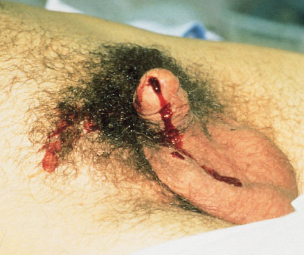
You’re seeing a trauma patient, probably a transfer from somewhere else. Either they told you there “may have been” some blood at the tip of the urethra, or maybe you see it smearing the outside of a urinary catheter that’s already in place! How do you proceed from here?

First, try not to get into that situation. Make sure that everyone on your team knows that gross blood at the meatus, male or female, means urethral injury until proven otherwise. If it’s not gross blood, it could be that the patient was incontinent and has hematuria from other causes. The fear with passing a catheter across a urethral injury is that it may convert a partial tear to a complete one. Reconstruction and complications from the latter are far more serious.

But the catheter is there. What to do?

First, leave the catheter in place. You must assume that the injury is present, and you need to rule it in or rule it out in order to decide what to do with the catheter. If the injury is not really there, then you can remove the catheter when indicated. If it really is present, then the urethral injury is being treated appropriately.

Next, do a urethrogram. I’ve previously described how to do it here, but the technique I describe is only appropriate for uncatheterized patients. The technique must be modified to use thin contrast and a method to inject alongside the catheter. To do this, fill a 20-30cc syringe with contrast (Ultravist or similar liquid) and put an 18 gauge IV catheter on the tip (no needles, please). Slide the IV catheter alongside the urinary catheter, clamp the meatus with your fingers, pull the penis to the side and inject under fluoroscopy. The contrast column will not be as vivid as with a regular urethrogram because it is outlining the urinary catheter, so there is less volume.

If the contrast travels the length of the urethra and enters the bladder without leaking out into soft tissue, there is no injury. If there is contrast leakage, stop injecting and plan to call a urologist.

Finally, be on the lookout for associated injuries. Urethral injuries are frequently found in patients with anterior pelvic fractures and perineal injuries.

Related post:

Link: blood at the urethral meatus (Atlas-Emerg-Medicine.org.ua from McGraw-Hill)

Thoughts On Traumatic Hematuria: Part 2

Yesterday, I discussed blood in the urine from a urethra. As I mentioned, there is typically not much from that particular injury. Today, I’ll dig into the three causes of real hematuria.

All of these tubes show gross hematuria except the one on the right.

  • Bladder injury. This can occur with either blunt or penetrating injury. The degree of hematuria is variable with stabs or gunshots, but tends to be much darker in blunt injury. This happens because the size of the bladder injury tends to be greater with blunt force. The bladder injury is not necessarily full-thickness with blunt trauma. It may just be some wall contusion and underlying mucosal injury. But frequently, with seat belt injury and/or A-P compression injuries to the pelvis (“open book”), the injury is full thickness.
    • Tip: If less than 50cc of very dark urine flow from the catheter upon insertion, it is likely that your patient has an intraperitoneal bladder rupture!
  • Ureteral injury. This injury is very rare. The most common mechanism is penetrating, but this structure is so small and deep that it seldom gets hit by naything. Patients with multiple lumbar transverse process fractures will occasionally have a small amount of hematuria, probably from a minor contusion. More often than not, the hematuria is microscopic, so we should never know about it.
  • Kidney injury. The most important fact regarding renal injury is that the degree of injury has no correlation with the amount of hematuria. The most devastating injury, a devascularized kidney, frequently has little if any gross hematuria. And conversely, a very minor contusion can produce very red urine.

So what about diagnosis? It’s easy! If you see gross hematuria, insert a foley catheter (if not already done) and order a CT of the abdomen/pelvis with contrast, as well as a CT cystogram. The latter must not be done using passive filling of the bladder with a clamped catheter. Contrast must be infused into the bladder under pressure to ensure a bladder injury can be identified.

CT scan is an excellent tool for defining injuries to kidney, ureter, and bladder, and will identify extravasation into specific places and allow grading. Specific management will be the topic of future posts.

Thoughts On Traumatic Hematuria: Part 1

I’ve seen a number of patients recently with bloody urine, and that is prompting me to provide some (written) clarity to others who need to manage this clinical problem. I’ll try to keep it organized!

There are two kinds of hematuria in trauma: blood that you can see with the naked eye, and…

Okay, so there’s only one. Trauma professionals do not care about microscopic hematuria. It does not change clinical management. Sure, your patient might have a renal contusion, but you won’t do anything about that. Or, he/she might have an infarcting kidney. And you can’t do anything about that. If you order a urinalysis, you might see a few RBCs. Don’t let this lead you down the path of looking for a source. You’ll end up ordering lots of tests and additional imaging, and generally will have nothing to show for it at the end. It’s not your job to spend good money on the very rare chance of finding something clinically significant.

Both of these specimens have blood in them. You can’t see it on the left, so don’t go looking for it with a microscope.

There are four sources of blood in the urine.

1. The first source does not generally cause hematuria, but can occasionally cause a few visible wisps of blood. That source is a urethral injury. The textbook teaching, and it’s good advice, is to look at the urethral meatus in your trauma patient, especially if you are contemplating insertion of a urinary catheter. If you see a few drops of blood, pause to consider. Sometimes, the blood is no longer visible, but might be present as a few well-placed drops on the patient’s underwear. So have a look at that, too, especially in patients with high risk injuries such as A-P compression pelvic fractures (think, lots of ramus fractures or pubic diastasis).

If you didn’t notice it and inserted the catheter anyway, you might see a few wisps of blood in the tubing as you place it. More often than not, this is just run of the mill irritation of the mucosa by the catheter, but always keep the possibility of an injury in mind.

Tomorrow, I’ll discuss the remaining three sources, and what to do about them.

Related posts: