I recently wrote about how the completion chest x-ray can lie after insertion of a chest tube. The chest x-ray image is a 2-D representation of the patient, but you really can’t tell where the tube lies in the third dimension (front to back). That’s how a trauma professional can get suckered into thinking they just put a perfect chest tube in, when in reality they have not.
How can you be sure of the position as you are putting it in? It’s a nuisance to have to reposition it after you’ve taken down your sterile field. Here are a few suggestions, but pay particular attention to the last one. I think it’s the best.
- Make the incision large enough so that you can visually confirm that the last hole is inside the thoracic cavity. This option is somewhat okay for thinner patients. But it leads to a larger than necessary incision, especially in patients who are obese. Not a great idea.
- Estimate proper depth before insertion. Hold the tube over the patient’s chest, and note the distance mark printed on the tube when the tip is placed halfway across the hemithorax (just medial to the nipple). This does take into account the amount of soft tissue on the lateral chest, but is not terribly accurate and you may accidentally contaminate the tube. The usual depth for a patient with normal body habitus is 12-14 cm at the skin. A better choice.
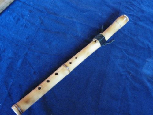
- Use the “bamboo flute” technique. Once you have entered the pleural space and placed the end of the tube into it, locate and place your finger firmly over the last hole, like you were playing a flute. Keep it there as you slide the tube in until your finger contacts the ribs around the insertion point. It should be at a right angle to the chest wall. Then push it in another 2-4 cm. As long as you have performed a nice dissection down to the chest wall, this technique is close to foolproof. And double-check by making sure that the tube is at least 12-14 cm at the skin. IMHO, this is the best technique.

This is not a chest tube!
Related posts:

