The Chance Fracture

Centers that take care of blunt trauma are familiar with the spectrum of injury that is directly attributable to seat belt use. Although proper restraint significantly decreases mortality and serious head injury, seat belts can cause visceral injury, especially to small bowel.

Lap belt use has been associated with Chance fracture (flexion distraction injury to the lumbar spine) since 1982. The association between seat belts and intra-abdominal injury, especially with an obvious “seat belt sign” was first described in 1987.
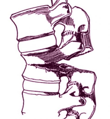
chance-fracture-21

Chance fracture. The vertebra appears to split in half from posterior to anterior.

Twenty years ago, orthopedic surgeons in Manitoba finally put two and two together and reported a series of 7 cases of Chance fractures. They noted that 6 of the fractures were associated with restraint use. Seat belt sign was also present in 5 of the 6 patients with fractures and three of the six had bowel injuries.

The authors noted that many provinces were mandating seatbelt use at the time, and they predicted that the number of Chance fractures, seat belt signs and hollow viscus injuries would increase. On the positive side, the number of deaths and serious head injuries would be expected to decline.

Although this was a small series, it finally cemented the unusual Chance fracture, seat belt sign, and bowel injury after motor vehicle trauma.

Thankfully, three point restraints (lap belt + shoulder harness) has been required in the seats next to doors for a long time. And since 2007, they have been mandated in the middle seat as well. Thus, these injuries seldom occur in any but the oldest (beater) cars on the road. They are seen more frequently now with sports and extreme sports injuries.

Chance fractures are frequently unstable, involving all three columns of the spine. The anterior column fails under compression, and the middle and posterior columns fail from the distraction mechanism. Usually, this fracture pattern requires operative fixation. However, if the posterior column is intact, a TLSO brace can be tried. This fracture is at risk for non-union and development of kyphosis or a flat back, which can lead to chronic pain and an abnormal posture.

Reference: Pediatric Chance Fractures: Association with Intra-abdominal Injuries and Seatbelt Use. Reid et al. J Trauma 30(4) 384-91, 1990.


Submental Intubation – The Video!

Yesterday, I described a technique for providing a secure yet short-term airway tailored to patients who can’t have a tube in their mouth or nose. Patients undergoing multiple facial fracture repair are probably the best candidates for this procedure.

A picture may be worth a thousand words, but a video is even better. Please note that it is explicit and shows the blow by blow surgical procedure. Of note, it is a quick and relatively simple advanced airway technique.

YouTube player

Related post:

By Request: Submental Intubation

I’ve had a number of recent requests regarding this technique, so I’m updating and reposting today and tomorrow.

Here’s one of the weirder procedures I’ve seen in some time. Imagine that you need a definitive airway, but you can’t use the face for some reason (mouth or nose). The usual choice would be a tracheostomy, right? But what if you only need it for a few days? Typically, once placed, trachs must be kept for a few weeks before decannulation is safe.

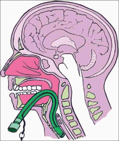
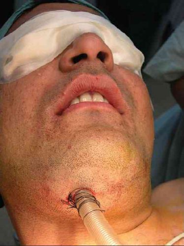
Enter submental intubation. This technique involves passing an endotracheal tube through the anterior floor of the mouth, and then down the airway. This leaves the facial bones, mandible, and skull base untouched.

tumblr_inline_mvds1c14ye1qa4rug1


The technique is straightforward:

  • After initially intubating the patient  orotracheally, a 1.5cm incision is created just off the midline in the submental area under the chin.
  • Using a hemostat, all layers are penetrated, entering the oropharynx just lateral to the tongue.
  • A 1.5cm incision is then made at the puncture site, parallel to the gum line of the lower teeth.
  • The ET tube is removed from the ventilator circuit, and the connector at the proximal end of the tube is removed.
  • The hemostat is placed through the chin incision again. The proximal end of the ET tube is curled into the oropharynx and grasped with the hemostat, then pulled out through the skin under the chin, leaving the distal (balloon) end in the trachea.
  • The connector is reinserted, and the tube is then hooked up to the anesthesia circuit again.
  • The tube is then secured using a stitch under the chin.

After a final position check, the surgical procedure can commence. Cool!


There are a number of variations on this technique, so you may encounter slightly different descriptions. The tube can be pulled at the end of the procedure, or left for a few days to ensure safe extubation, if needed.

tumblr_inline_mvdsm0zd8b1qa4rug1

A small series of 10 patients undergoing this technique was reviewed, and there were no short or long term problems. Scarring under the chin was acceptable, and was probably less noticeable than a trach scar.

Bottom line: This is a unique and creative method for intubating patients with very short-term airway needs while their facial fractures are being fixed. Brilliant idea!

Tomorrow: Submental intubation – the video!

Reference: Submental intubation in patients with panfacial fractures: a prospective study. Indian J Anaesth 55(3):299-304, 2011.

Photo source: internet

Help! My Consultant Won’t Come In To See A Patient!

Consultants provide very important services to trauma patients in the ED and inpatient settings. The trauma professionals managing those patients can’t know everything (although we sometimes think we do). But occasionally our patients present issues that require evaluation by other experts in order to guarantee excellent care.

Sometimes our consultants want to do too much, or make recommendations that are not really in their area of expertise (e.g. a cardiologist evaluating a cardiac contusion). See the related post link below for tips on this situation.

But sometimes you know what the patient needs, but the consultant doesn’t agree or doesn’t do what you expect. Or they don’t want to come in when called. What to do?

Here are some tips:

The patient is in the ED and the consultant won’t come in to see the patient.

  • Are they right? Does that problem really need to be dealt with in the ED in the middle of the night? Many simple fractures and wounds do not need immediate attention. They can be dressed/splinted, the patient reassured, and instructed to see the consultant in the clinic the next day.
  • Is your knowledge of current management of the condition correct? Perhaps it has evolved, and it is now commonplace to temporize and deal with the problem as an outpatient during business hours. Make sure you are up on the current literature.

The patient is in the ED and the consultant won’t come in to see the patient, and you are sure that they should! Now what?

  • Call them personally (not a resident, midlevel provider, or any other intermediary) and clearly and concisely explain the situation, and your assessment of why the problem needs their immediate attention.
  • Listen to or elicit their rationale for not seeing the patient. If legitimate, this may help educate you and modify your future management of similar patients. If the rationale is not legitimate, inform them (tactfully) that this is at odds with your education/training/experience with other providers. Ask them to further explain, if they can.

If they still won’t come in despite what you think is a legitimate need, then you must calculate a quick risk:benefit ratio. Will any patient harm occur if the consultant does not see the patient? And what is the professional damage that you will incur if you move on to the next steps. If you believe that harm will occur, here are your options, from least to most damaging to your professional status at the hospital:

  • Contact another consultant in the same or overlapping specialty (if there is one). Apologize for the fact that you know they are not on call, and explain the situation.
  • Appeal to a higher authority. Contact the trauma medical director, service chief, or hospital administrator and see if they can intervene.
  • Explain to the consultant that you truly believe that harm will occur, and you will have to document that fact in the medical record as well as their failure to respond. In some cases, this will shake them loose, but they will certainly be pissed.
  • If all else fails, see if you can find a service that will help you by accepting the patient as an admission so they can be managed appropriately the next day. But then follow through by reporting the event to appropriate people including chief of staff, chief medical officer, VPMA, hospital quality department, and risk management. This is the nuclear option, so be prepared for the fallout.

Bottom line: This is not a fun situation to find yourself in. Good luck!

Related post:

Retained Foreign Objects After Penetrating Injury

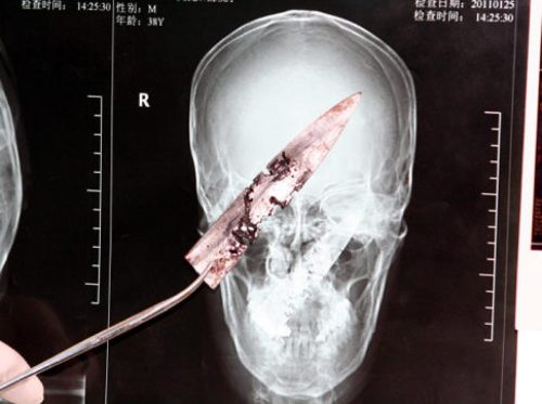
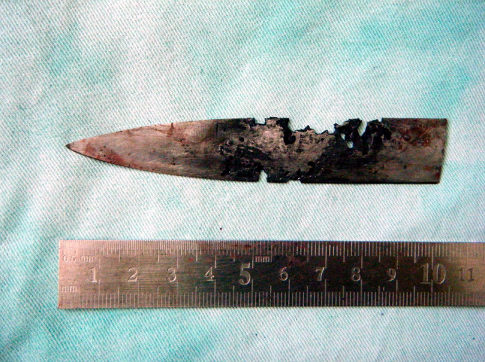
A Chinese man was in the news a few years back after having a four inch knife blade removed from his head. It had been there for four years!  The knife blade broke off after he had been stabbed under the chin. Unfortunately, he was unaware that any part of the knife had been retained. It remained partly within the nasopharynx and the tip came to rest behind his left eye. His symptoms included headaches, stuffy nose and bad breath. The picture below shows the badly corroded blade in front of some of his radiographic images.

See the video at the bottom of this post for more details and images.

knife-in-head

What is the best way to deal with a problem like this? Here are some practical tips:

First, get in the habit of imaging any body part with a penetrating injury. Retained objects can be as simple as gravel or as complicated as the knife blade above. And remember, some patients who have been stabbed present with a simple laceration but don’t want to tell you how they got it. Image before you close it!

Next, don’t remove it. This is common knowledge, but innocent looking objects (pencils, nails) can penetrate arteries and keep them from bleeding while embedded. Unpleasant and sometimes fatal bleeding can ensue if pulled out.

If you do not have specialists versed in the body regions involved in the injury, transfer immediately with the object secured in place. For objects penetrating minimally complex areas like the extremities, surgeons may opt to carefully remove it in the emergency department, or may elect to do so in the operating room.

Injuries to complex areas should undergo high resolution CT scanning so that 3D reconstruction can be performed if needed. The surgical specialists can then plan the operative approach. This is dictated by the anatomy of the area(s) involved and the architecture of the object (think about hooks and barbs). For objects located near critical areas, an operative exposure must be selected that provides access to all portions of it, and allows for rapid vascular control if needed.

YouTube player