IV Contrast and Trauma

We use CT scanning in trauma care so much that we tend to take it (and its safety) for granted. I’ve written quite a bit about thoughtful use of radiographic studies to achieve a reasonable patient exposure to xrays. But another thing to think about is the use of IV contrast.

IV contrast is a hyperosmolar solution that contains some substance (usually an iodine compound) that is radiopaque to some degree. It has been shown to have a significant impact on short-term kidney function and in some cases can cause renal failure.

Here are some facts you need to know:

  • Contrast nephrotoxicity is defined as a 25% increase in serum creatinine, usually within the first 3 days after administration
  • There is usually normal urine output and minimal to no proteinuria
  • In most cases, renal function returns to normal after 3-4 days
  • Nephrotoxicity almost never occurs in people with normal baseline kidney function
  • Large or repeated doses given within 72 hours greatly increase risk for toxicity
  • Old age and pre-existing diabetic renal impairment also greatly increase risk

If you must give contrast to a patient who is at risk, make sure they are volume expanded (tough in trauma patients), or consider giving acetylcysteine or using isosmolar contrast (controversial, may still cause toxicity).

Bottom line: If you are considering contrast CT, try to get a history to see if the patient is at risk for nephrotoxicity. Also consider all of the studies that will be needed and try to consolidate your contrast dosing. For example, you can get CT chest/abdomen/pelvis and CT angio of the neck with one contrast bolus. Consider low dose contrast injection if the patient needs formal angiographic studies in the IR suite. Always think about the global needs of your patient and plan accordingly (and safely).

Reference: Contrast media and the kidney. British J Radiol 76:513-518, 2003.

Torso Trauma CT (Nearly) ALWAYS Requires Contrast

Most stable patients with blunt trauma undergo CT scanning these days. Hopefully, it’s done thoughtfully to optimize the risk/benefit ratio using a well-designed imaging protocol. The majority of these torso imaging protocols call for the use of IV contrast. But as I’ve written before, this can pose risks, especially to the elderly and others who have some degree of renal impairment.

Unfortunately, I occasionally encounter scans done at other hospitals that omit the use of contrast. This usually hinders diagnosis significantly. And it’s usually not clear why this happened, so let’s think about it a bit.

The use of contrast in CT is designed to show blood, or things that are filled with lots of blood. Specifically, a great deal of detail about the blood vessels and solid organs is displayed.

Let’s break it down by type of scan:

  • Chest – we are really only interested in the aorta. The only way to reliably demonstrate an aortic injury is by using contrast. And this is one of those injuries that, if you miss it, the patient is very likely to die from it. Therefore, if you are ordering a chest CT properly, you must add contrast.
  • Abdomen/pelvis – generally, we are looking for solid organ injury, potential mesenteric injuries, and extravasation of blood from organs or soft tissue. Once again, the only way to really see any of these is with contrast enhancement.
  • Vascular – CT is replacing conventional angiography for the investigation of vascular injury in many cases. Obviously, this study is worthless without the contrast.

Bottom line: Pretty much any CT of the chest, blood vessels, or abdomen/pelvis must have IV contrast injected for accurate diagnosis. But what if your patient is old, or is known to have some degree of renal impairment? First, decide if you can wait until a point of care or standard creatinine measurement is done. If you can, use the result to do your own risk/benefit calculation. Is the injury you are worried about potentially life-threatening AND reasonably likely? Are there other less harmful ways to detect it? Then use them. And if you really do need the study in a patient with renal dysfunction, give the contrast, monitor the serum creatinine regularly, and do what you can to optimize and protect their renal function over the next several days.

Once Again: Leg Fasciotomy With Less Than Two Incisions

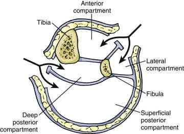
It seems like this topic keeps on coming up! This is the second article I’ve seen this year that describes a variation on the single-incision leg fasciotomy. In the classic two-incision approach, the lateral incision gives access to the anterior and lateral compartments, and the medial incision to the posterior and deep posterior. See below.

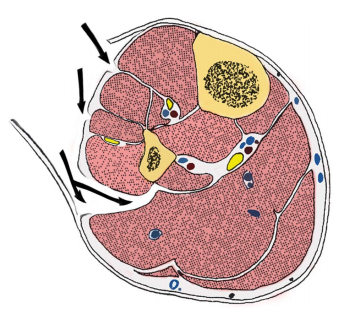
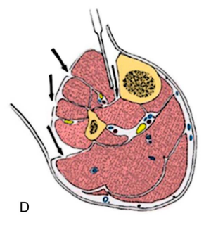
The more “standard” single-incision approaches either go through (i.e. removes part of) or around, the fibula. In the diagram below, the arrows point to the access points into the anterior, lateral, posterior and deep posterior from top to bottom.

In the “new” variation described, the authors slide along the lateral edge of the tibia to get to the deep posterior compartment.

This approach requires stripping the tibialis

anterior muscle away from the tibia, which some orthopods may argue interferes with healing. And, as with the other single-incision technique, the procedure may take additional time.

Bottom line: I’m still not a big fan of single-incision fasciotomy. My main reason is that most surgeons are not as familiar with the technique. And patients who have a potentially limb threatening process are not the best to learn on. I have seen too many incomplete fasciotomies with persistent compartment syndrome in my career. 

So unless you are being mentored by someone who is well versed in the technique, use the two incision technique and use a cadaver to practice your single incision operation.

Reference:  A Single-Incision Fasciotomy for Compartment Syndrome
of the Lower Leg. J Ortho Surg 30(7):e252-e255, 2016.

The Lead Gown Pull-Up: Part 2

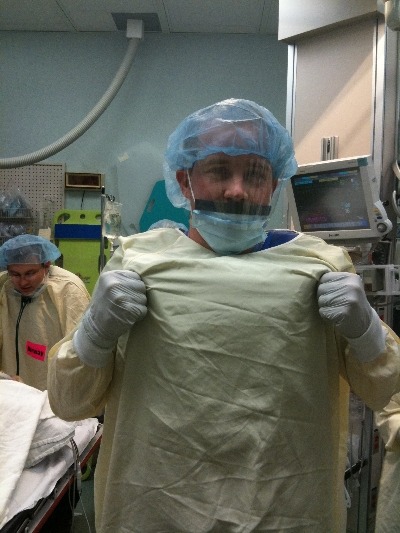
Okay, so you’ve seen “other people” wearing perfectly good lead aprons lifting them up to their chin during portable x-rays in the trauma bay. Is that really necessary, or is it just an urban legend?

After hitting the medical radiation physics books (really light reading, I must say), I’ve finally got an answer. Let’s say that the xray is taken in the “usual fashion”:

  • Tube is approximately 5 feet above the xray plate
  • Typical chest settings of 85kVp, 2mAs, 3mm Al filtration
  • Xray plate is 35x43cm

The calculated exposure to the patient is 52 microGrays. Most of the radiation goes through the patient onto the plate. A very small amount reflects off their bones and the table itself. This is the scatter we worry about.

So let’s assume that the closest person to the patient is 3 feet away. Remember that radiation intensity diminishes as the square of the distance. So if the distance doubles, the intensity decreases to one fourth. By calculating the intensity of the small amount of scatter at 3 feet from the patient, we come up with a whopping 0.2 microGrays. Since most people are even further away, the dose is much, much less for them.

Let’s put it perspective now. The background radiation we are exposed to every day (from cosmic rays, brick buildings, etc) amounts to about 2400 microGrays per year. So 0.2 microGrays from chest x-ray scatter is less than the radiation we are exposed to naturally every hour!

The bottom line: unless you need to work out you shoulders and pecs, you probably don’t bother to lift your lead apron every time the portable x-ray unit beeps. It’s a waste of time and effort! Just stand back and enjoy!