Now Where Did That Bullet Go? The Answer!

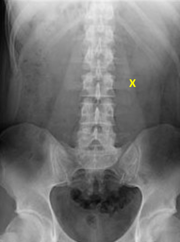
Previously, I presented a scenario where a victim of a gunshot to the abdomen was taken to the OR after obtaining the image below. No bullet was seen on the x-ray, and none was found at the time operation.

image

Where could it have gone? Let’s assume that the surgeon did a good job, and it is not in the abdomen. Any more. There are several possibilities.

Does the x-ray cover the correct area? To cover a straighforward abdominal gunshot, it needs to show diaphragms to perineum, side to side. In these days of “super size me”, that usually doesn’t happen with one image. Look carefully at the one above. It doesn’t show any portion of the diaphragm, and doesn’t go low enough, either. And although the right flank can be seen, the left is cut off. So in this case, the bullet could be in the soft tissues of the torso, in the extraperitoneal rectal area, or near the diaphragm in the liver.

It could have moved outside the area of the initial x-ray. The most common mechanism for this is entry into the vascular system. If it enters the venous system, it will end up in the heart or pulmonary artery somewhere. This will be obvious when you get a chest x-ray. If it enters the aorta, it will embolize into the lower extremities. This fact should be painfully obvious when you check the pulses in the lower extremities

The patient could poop it out if it entered their GI tract. This could happen if you wait to get additional images of the abdomen. If you bracket it with x-rays immediately, this should not happen. 

In theory, the bullet could enter the bladder and get urinated out. This won’t happen if a catheter is in place. And it’s probably unrealistic because most bullets would cause tremendous pain passing, and would probably obstruct the urethra anyway.

Finally, it could have bounced. Never count on this one. Bullets can and do enter partially, then stop or fall out. They can cause underlying perforation of the peritoneum, and they can bruise nearby structures. This is extremely uncommon and should be a diagnosis of last resort!

Bottom line: If patient condition permits, the patient with a gunshot to the abdomen who will be taken to the OR should have any wounds marked and an initial abdominal image obtained that shows the entire abdomen. This may take multiple attempts. The image can be very helpful in directing the exploration and finding wounds. If it is not seen on the initial image(s), check the lower extremity pulses and obtain a chest x-ray to locate the bullet prior to the case.

Now Where Did That Bullet Go?

Here’s an interesting case for you to consider. A male victim of an assault is brought to your emergency department with a gunshot to the abdomen. He is met by your team as a trauma activation. Vitals are stable, but he has guarding and rigidity. A single abdominal x-ray is obtained, and then the patient is taken off to surgery. I’ve marked the entry site on the anterior abdominal wall below. There is no exit wound.

In the operating room a laparotomy is performed. There is a hole in the fascia under the skin penetration. A small hematoma is seen in the underlying transverse mesocolon, well away from the bowel itself. An extensive search is carried out, but no other holes, injuries, or evidence of a bullet is found. Ultimately, the abdomen is closed and the patient is admitted to a ward bed.

WTF? Where did the bullet go? What do you think the possibilities are? Please leave comments today either here or on Twitter. I’ll analyze this puzzling situation tomorrow!

Disclosure for my social media compliance police: this patient was not treated at Regions Hospital, and the x-ray was obtained and modified from the internet.

Prehospital Lift-Assist Calls

Here’s something I was completely unaware of until just a few years ago. A number of 9-1-1 calls (quite a few, I am told) are made, not for injury or illness, but because the caller needs help getting back into bed, chair, etc. It is also common that prehospital providers are frequently called back to the same location for the same problem, or a more serious one, within hours or days.

Yet another study from Yale looked at the details of lift-assist calls in one city in Connecticut (population 29,000) during a 6 year period. The town has a fire department based EMS system with both basic and advanced life support, and they respond to 4,000 EMS calls per year.

Here are the factoids:

  • Average crew time was about 20 minutes
  • 10% of cases required additional fire department equipment, either for forced entry or for assistance with bariatric patients
  • About 5% of all calls were for lift-assist, involving 535 addresses
  • Two thirds of all calls went to one third of those addresses (174 addresses)
  • There were 563 return calls to the same address within 30 days (usual age ~ 80)
  • Return calls were for another lift-assist (39%), a fall (8%), or an illness (47%)

Bottom line: It looks to me that we are not doing our elderly patients any favors by picking them up and putting them back in their chair/bed. Lift-assist calls are really a sentinel event for someone that is getting sick or who has crossed the threshold from being able to live independently to someone who needs a little more help (assisted living, etc). Prehospital personnel should systematically look at and report the home environment, and communities should automatically involve social services to help ensure the health and well being of the elder. And a second call to the same location should mandate a medical evaluation in an ED before return to the home.

Reference: A descriptive study of the “lift-assist” call. Prehospital Emergency Care 17(1):51-56, 2013.

The Lucas CPR Device And Pregnancy

A reader posed an interesting question last week: can you use the Lucas chest compression device in a pregnant patient?

The official company answer is “no.” Obviously, this is one those areas that is tough to get research approval on, and the number of pregnant patients who might need it is very small. So basically, we have little experience to go on.

That being said, the reality is that prehospital agencies can and do use it for these patients on occasion. There is only one published case report that I could find (see reference below). The thing that makes using this device a little more challenging is that, to optimize blood pressure, late term pregnant patients need to have the uterus rolled off of the vena cava. This means tipping the patient to her left. 

As you can see from the picture above, the design of the Lucas makes this a bit difficult. However, it can be done, either by tipping the board the patient is on or wedging something under the right side of the back plate.

And as always, make sure that you adhere to your local policies and procedures, or have permission from your medical director to use this device in this particular situation.

Reference: Cardiac arrest and resuscitation with an automatic mechanical chest compression device (LUCAS) due to anaphylaxis of a woman receiving caesarean section because of pre-eclampsia. Resuscitation 68(1):155-159, 2005.

Autotransfusing Blood Lost Through The Chest Tube

Autotransfusing blood that has been shed from the chest tube is an easy way to resuscitate trauma patients with significant hemorrhage from the chest. Plus, it’s usually not contaminated from bowel injury and it doesn’t need any fancy equipment to prepare it for infusion.

It looks like fresh whole blood in the collection system. But is it? A prospective study of 22 patients was carried out to answer this question. A blood sample from the collection system of trauma patients with more than 50 cc of blood loss in 4 hours was analyzed for hematology, electrolyte and coagulation profiles.

Here are the factoids:

  • The hemoglobin and hematocrit from the chest tube were lower than venous blood (Hgb by about 2 grams, Hct by 7.5%)
  • Platelet count was very low in chest tube blood
  • Potassium was higher (4.9 mmol/L), but not dangerously so
  • INR, PTT, TT, Factor V and fibrinogen were unmeasurable

Bottom line: Although shed blood from the chest looks like whole blood, it’s missing key coagulation factors and will not clot. Reinfusing it will boost oxygen carrying capacity, but it won’t help with clotting. You may use it as part of your massive transfusion protocol, but don’t forget to give plasma and platelets according to protocol. This also explains why you don’t need to add an anticoagulant to the autotransfusion unit prior to collecting or giving the shed blood!

Related post: Chest tubes and autotransfusion

Reference: Autotransfusion of hemothorax blood in trauma patients: is it the same as fresh whole blood? Am J Surg 202(6):817-822, 2011.