Bowel Sounds, Or Just Plain BS?

“Bowel sounds are normal”

How often do you see this on an H&P? Probably a lot more often than they are actually listened for, I would wager. But what do they really mean? Are they important to trauma professionals?

(Un)fortunately, there’s not a whole lot of research that’s looked at this mundane item. And pretty much all of it deals with surgical pathology (e.g. SBO) or the state of the postop abdomen. Over the years, papers have been published about the basics, and I will summarize them below:

  • Where to listen? Traditionally, auscultation is carried out in all four abdominal quadrants. However, sound transmission is such that listening centrally is usually sufficient.
  • Listen before palpation? Some papers suggest that palpation may stimulate peristalsis, so you should listen first.
  • How long should you listen? Reports vary from 30 seconds to 7 minutes (!)
  • Significance? This is the big question. We’re not expecting to find hyperactive or high pitched sounds suggestive of surgical pathology here. Really, we’re just looking for sounds or no sounds.

But does it make a difference whether we hear anything or not?

Bottom line: In trauma, we don’t care about BS! We’ve all had patients with minimal injury and no bowel sounds, as well as patients with severe abdominal injury and normal ones. We certainly don’t have time to spend several minutes listening for something that has no bearing on our clinical assessment of the patient. Skip this unnecessary part of the physical exam, and continue on with your real evaluation!

Reference: A critical review of auscultating bowel sounds. Br J Nursing 18(18):1125-1129, 2009.

Is This A Good Chest Tube? The Answer!

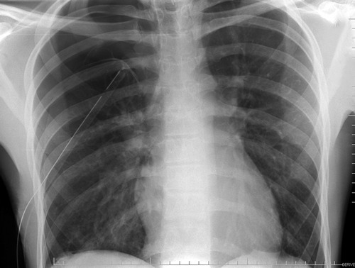
I showed you this x-ray the other day, and asked what the problem was.

If you look carefully, you can see the lung outline in the middle of the right side of the chest. Big-time pneumothorax despite what looks like a perfectly placed tube. There are several possible explanations, and many of you sent me your guesses:

  • The tube is in the lung. This rarely happens to normal lungs. Sure, you can probably do it to an ARDS lung, but otherwise it’s not likely.
  • The tube is in the fissure. This does happen on occasion, but not often. And many times it works anyway.
  • The tube is occluded or kinked. A PA or AP chest x-ray will show the kink, although bent tubes frequently work anyway. If a hemothorax is present, it is possible that a clot is plugging the tube. Clearing a plugged tube will be the subject of another post.
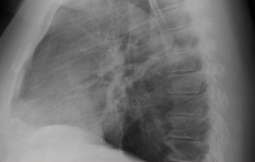
  • It’s not really a chest tube. Hopefully, this would have been detected when it was placed, but it’s not always. The chest x-ray above looks great, right? Unfortunately, it’s a 2 dimensional representation of a 3-D object. Where is that tube in the z-axis?

In this case the correct answer is the last one. This is one time when I would actually recommend a lateral chest x-ray. Have a look at the result. You can clearly see the tube snaking around into the soft tissues of the back.


Bottom line: Remember that a perfect x-ray doesn’t necessarily mean a perfect tube. Go through the various possibilities quickly, and make it work.

Related posts:

The Trauma Professional’s Blog Is 5 Years Old!

I know, it’s hard to believe, but this blog turns 5 years old today! 

It’s been so much fun to see my readership grow over the past years. I’m so thankful to my thousands of readers all over the world. I’m also grateful for all the questions that I get from trainees and trauma professionals everywhere. It keeps me on my toes and keeps me reading and learning every day.

In the coming year, I will continue to try to provide answers to those pesky questions that you really can’t find anywhere else. I’ll also continue to try to digest stuff for you that is in the literature and give you my take on it, as well as whether I think it’s important or just plain bogus.

So THANK YOU very much, and have a fantastic new year. Keep those questions coming so I’ve got plenty to write about in the coming year.

Michael