What The Heck? Bicycle Trauma Part 2

Yesterday’s puzzle involved a young male who drove a handlebar into his abdomen. Little additional information was given, other than one slice of his abdominal CT scan. So what’s the problem?

The textbooks always associate handlebars with pancreatic and duodenal injuries, and these should always be looked for. However, the scan slice in this case was taken lower, within the pelvis. Too low to show you either of those organs.

As I’ve said before, be systematic when reading xray images. We automatically focus on the viscera and bones. Look at those areas, make sure you can identify each structure that you see, and look for any anomalies. 

But don’t forget the soft tissue! In this case, the child doesn’t have much. Take a closer look at the same slice and see if you can figure it out by tomorrow.

ICP Monitoring: Less Is More?

Management of severe traumatic brain injury (TBI) routinely involves monitoring and control of cerebral perfusion pressure. Monitoring is typically accomplished with an invasive monitor, with the extraventricular drain (EVD) and fiberoptic intraparenchymal monitors (IP) being the most common.

The extraventricular drain is preferred in many centers because it not only monitors pressures, but it can also be used to drain cerebrospinal fluid (CSF) to actively try to decrease intracranial pressure (ICP). But could less really be more? Surgeons at Massachusetts General reviewed 229 patients with one of these monitors, looking at outcomes and complications. They found the following interesting results:

  • There was no difference in mortality between the two monitor types
  • The EVD patients did not require surgical decompression as often, possibly because of the ability to decrease ICP through drainage
  • The EVD patients were monitored longer, and had a longer ICU length of stay. This was also associated with a longer hospital length of stay.
  • Complications were much more common in the extraventricular drain group (31%). The most common complications were no drainage / thrombosis (15%) and malposition (10%). Hemorrhage only occurred in 1.6% of patients. 
  • Fiberoptic monitors had a lower complication rate (8%). The most common was malfunction leading to loss of monitoring (12%). Hemorrhage only occurred in 0.6% of patients.

Bottom line: Don’t change your monitoring technique yet. Much more work needs to be done to flesh out this small retrospective study. But it should prompt us to take a critical look for better indications and contraindications for each type of monitor.

Reference: Intraparenchymal versus extracranial ventricular drain intracranial pressure monitors in traumatic brain injury: less is more?J Am Coll Surg 214(6):950-957, 2012.

Family Presence During Trauma Resuscitation

There’s a lot of talk in trauma nursing circles about family presence during trauma resuscitation (FPDR). But after searching far and wide, I have not been able to find much literature about it. There are a few papers about family reaction to it, and a few more about healthcare providers’ reactions. But the science is not yet very good.

Typical arguments against it from (mainly) doctors and some nurses, and my rebuttals, are as follows:

  • Family members will think the trauma resuscitation is chaoticthen fix your trauma team; you’ve got a problem
  • They will slow the team down – an analysis has shown no such effect
  • Family will be traumatized by seeing what we do – family members have seen simulations on TV, so they have a pretty good idea of what’s going on
  • The doctors and hospital are more likely to be sued – actually, this is probably less likely, because the family has actually seen that you’ve done everything possible for their loved one

Personally, I’m very much for it, especially in the pediatric age group. I encourage all trauma programs to develop a policy to enable FPDR. Here are some key pointers:

  • Only allow one key family member in the resuscitation room. Have them decide who it will be. This limits confusion and congestion.
  • Assign a “medical interpreter” to stand with them in the room, preferably a nurse. The role of this person is twofold: to explain what is being done and why, and to make sure that they remain safe. If they have a hard time coping, appear to be getting faint, or misbehave in any way, it’s time for them to leave.
  • Involve the family member as much as practical. Have them stand near the patient’s head so they can communicate with them, or at least see them.
  • Keep the trauma team organized and professional. It’s been my experience that having family in the room puts everybody on their best behavior.

Bottom line: Surveys have shown that family members tend to be more satisfied with care and more convinced that everything possible was done if they are able to witness what may be the final moments of their loved one’s life. A little planning goes a long way in allowing FPDR in your emergency department.

References:

  • Health care providers’ attitudes regarding family presence during resuscitation of adults: an integrated review of the literature. Clin Nurse Spec 24(3):161-174, 2010.
  • Attitudes of healthcare staff and patients’ family members towards family presence during resuscitation in adult critical care units. J Clin Nurs. 21(13-14):2083-2093, 2012.
  • Family presence during pediatric trauma team activation: an assessment of a structured program. Pediatrics 120(3):e565-574, 2007.

CT Scanning After Gunshot To The Abdomen

Yesterday’s question related to ordering a CT scan in patients with a gunshot to the abdomen. Should it ever be needed? In general, if you encounter this question on an exam, the answer should be no. However, medicine in general and trauma in particular are not so black and white. There are always exceptions to the rules. 

Generally speaking, gunshots to the abdomen have a 90+ percent chance of causing an injury that requires repair. Pretty good odds that the patient needs a laparotomy. However, there are a few cases where further diagnosis may be okay.

In general, additional imaging is warranted if it will change the decision-making process in some way. In most gunshots to the abdomen, decision-making is very straightforward, and the patient must go to the OR without delay.

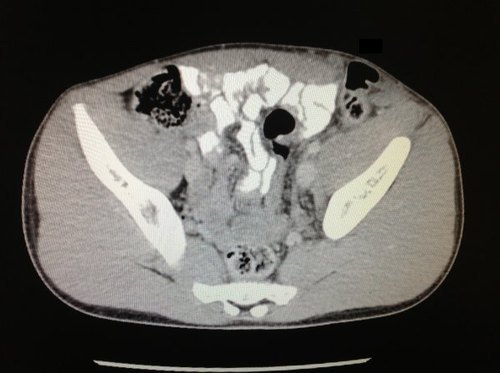
In some cases, there is a question as to whether or not the patient even needs an operation. The most common situation occurs when the wound could be tangential and completely extraperitoneal. These patients must be hemodynamically stable and without diffuse abdominal pain or tenderness to be considered for CT. Symptoms over the wound tract are acceptable. CT can show very clearly that the bullet stayed away from critical internal structures. These patients may even be discharged if they have no other injuries.

The other case is applicable in select patients with an obvious need for OR and who are hemodynamically stable. If a roadmap provided by CT would potentially cause the surgeon to limit, focus or expand the exploration, the scan may be justifiable. Most commonly, this occurs in patients with multiple gunshots, in whom the exact trajectories can’t be fully appreciated by looking at the holes and the known bullets seen on plain abdominal images.

Bottom line: CT scan in patients with gunshots to the abdomen should be a rare occurrence. There must be specific indications, and the patient must be hemodynamically stable. If the result may change the procedure in some way, it may be justifiable. Just be ready to explain your rationale to your trauma medical director! They will ask!