Trauma PI: Chasing Rumors

Trauma performance improvement (PI) is part art and part science. I tend to segregate the process into 3 segments: inputs, processing, and outputs. There are lots of possible inputs, including violation of specific audit filters (too long to OR, open fracture delay, etc.),  referrals from M&M discussions, incident reports and video reviews of trauma resuscitations, to name a few.

There is one PI input that has the potential to be a problem, though: word of mouth. You know, someone tells the trauma program manager that things just didn’t go well during that last trauma resuscitation. This is a perfectly legitimate way to identify PI issues. However, “word of mouth” can be categorized by source into “identified” and “anonymous." 

Word of mouth sources that are identified are not a problem. Anonymous ones are. All too often, these unsigned notes or suggestion box drops or phone messages are initiated by someone with an axe to grind. Most of the time, there is no basis for the incident that has been reported. The PI program can spend lots of time and energy trying to track down these perceived "problems”, and nothing ever comes of it.

There are two major problems with unsourced word of mouth “tips”:

  • There is no way to get additional information about the event from the source
  • It is not possible to thank the source for the information and let them know what was done to correct the issue

Bottom line: Performance improvement “tips” from anonymous sources are usually unfounded and a waste of time to investigate. Let it be known that your PI program is happy to receive written or verbal notices of potential problems that need to be pursued. However, every request must have a name and contact number and/or email included or it will be discarded.

Related posts:

CMS Meaningful Use And The Trauma Flow Sheet

Apologies to all my international readers. This US specific post may make your head spin.

Over the past two years, I’ve discussed the problems associated with trying to force the trauma flow sheet into an electronic form (see links below). It just doesn’t work yet. In 2009, the US government Centers for Medicare & Medicare Services (CMS) designed a set of financial incentives to move healthcare providers and hospitals toward using an electronic health record (EHR). This incentive program is called “meaningful use." Could this force trauma centers to use it for their trauma flow sheet?

The answer is a resounding NO. Meaningful Use seeks to reduce errors, improve the availability of patient information for providers, help develop best practices, and automate processes such as filling prescriptions. Hospitals have until the end of 2012 to complete Stage 1, in which they establish a baseline for EHR use. Stages 2 and 3 will follow in 2013 and 2015, where EHR usage will be further integrated by expanding the usage requirements. The exact guidelines for Stages 2 and 3 have not been determined yet.

So what is actually required in Stage 1? A list of 24 objectives (for hospitals) as been developed. A slightly different list has been developed for healthcare providers. The hospital list consists of 14 core objectives which must be met, and another list of 10 objectives, and the hospital can pick any 5 to meet.

Here is a summary of the core list. All must be implemented:

  • Use computerized order entry
  • Perform drug allergy and interaction checks
  • Maintain an active problem list
  • Maintain an active drug list
  • Maintain an active drug allergy list
  • Record certain demographic info
  • Record certain vital signs
  • Record smoking for patients age > 13
  • Report required clinical quality measures to CMS
  • Implement one clinical decision support rule
  • Provide an electronic copy of the patient’s health information if requested
  • Provide an electronic copy of any discharge instructions
  • Be able to exchange key health information between providers
  • Protect all of this EHR information

Here’s the non-core list. Any five of 10 must be implemented:

  • Implement drug formulary checks
  • Record advance directives
  • Incorporate lab tests
  • Be able to generate lists of patients
  • Identify patient education resources
  • Perform medication reconciliation
  • Help transition the patient to another setting of care
  • Submit data to immunization registries
  • Be able to report data to state registries
  • Be able to submit surveillance data to public health systems

Bottom line: Did you see any mention of the trauma flow sheet here? NO. There is no language requiring specific portions of the record to be in electronic form. And since technology still has not advanced to the point where meaningful data input or report output can be generated by an EHR version of the trauma flow sheet, it should not be used for this purpose. A paper trauma flow sheet that has been scanned into the EHR is still the gold standard.

Related posts:

Links:

Why Are We So Afraid Of IV Vitamin K?

Vitamin K has gotten a bad rap for years. There have been rumors of anaphylactic reactions to IV administration since I can remember. In the very old days, it was discovered that the vehicle used to deliver it (a variant of castor oil) was the likely culprit. But even after the vehicle was changed, rumors persisted. This led to the recommendation that Vitamin K be given subq or orally back in the 1990s. Even after 20 years, the reluctance to give it IV is still there.

In trauma, we need things fast. If someone has critical bleeding because they are taking warfarin, we want to begin reversing it quickly. And although the desired effects of Vitamin K may take hours to achieve, oral and subq absorption in trauma patients can be variable. IV is usually the way to go. So is IV Vitamin K such a bad thing?

A number of analyses and meta-analyses have been completed on this topic. A fairly comprehensive one looked at the literature over 30 years, and also reviewed the FDA’s adverse reaction database. The literature review found 23 cases of anaphylactic reaction, 3 of which were fatal. The FDA database uncovered 132 patients over a 30 year period, of which 24 were fatal.

Sounds bad, right? Remember, that’s 1 case per year in the literature and 4 per year in the FDA database. Now let’s put it into perspective. The overall incidence of anaphylaxis in the US is about 50 to 2000 episodes per 100,000, with about 1500 deaths annually. Induction of anesthesia results in about 10-15 anaphylactic events per 100,000, with the majority from neuromuscular blockers. 

Bottom line: Anaphylaxis is possible with anything that we ingest or inject! Anaphylaxis after IV Vitamin K administration is rare, estimated to be about 4 per 100,000. This is incredibly low. Many pharmacies stipulate that it be given by slow IV push (over 30 minutes), but anaphylaxis has been seen at lower doses and even with slow IV push. If your patient needs Vitamin K, don’t be reluctant to give it intravenously. Your patient is more likely to have a reaction from latex than from this medication. As with any drug, though, always monitor your patient for adverse reactions. 

References:

  • Anaphylactoid reactions to Vitamin K. J Thromb Thrombolysis 11(2):175-183, 2001.
  • Epidemiology of anaphylaxis: findings of the American College of Allergy, Asthma and Immunology Epidemiology of Anaphylaxis Working Group. Ann Allergy Asthma Innunol 97(5):596-602, 2006.

Temporal Bone Fracture

The temporal bone is a very complicated and important piece of bone located at the base of the skull. It houses a number of structures vital to hearing and balance, and is a conduit for several important nerves. Most fractures are due to blunt trauma, and although several classification systems exist, the majority are irregularly shaped and don’t lend themselves to easy categorization.

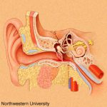
Clinically, a t-bone fracture should be suspected anytime that blood is seen in the ear canal or behind the eardrum after blunt head trauma. Patients may complain of dizziness or hearing loss. Examination may demonstrate some nystagmus in some cases. Most of the time, the diagnosis will not be made until a head CT is performed. The diagram above shows why this fracture can cause hearing problems (middle ear) or dizziness (inner ear).

There is usually no specific surgical intervention needed for this fracture. However, two important functional exams must be completed once diagnosed: facial nerve function and hearing. The facial nerve exam should document whether only portions of the nerve are affected vs all branches, and whether there is at least partial function. In head injured patients that can’t follow commands, even a grimace can demonstrate some function.

The ideal hearing test is carried out with a tuning fork. However, this is not usually convenient in acute trauma patients. Whispering and rubbing fingers near the patient’s ear can be used as a quick and dirty test. Awake patients will be able to tell you if their hearing is decreased.

Bottom line: Suspect temporal bone injuries in patients with any signs of basilar skull fracture. If the diagnosis is suspected or confirmed by CT scan, document the best facial nerve and hearing exam that you can. Contact your facial surgeon once the diagnosis is confirmed, and call them urgently if there is any loss of facial nerve function. Thin cut CT scans of the temporal bone are generally not necessary, and should not be ordered automatically unless the facial surgeon needs it for a specific reason.

Pneumothorax And Oxygen: The Final Post!

Okay, this is the last time I’m going to write about this. Hopefully I can provide the final nail in the coffin for this idea. Previously, the oldest paper I could find that was cited as a reason to use high inspired oxygen to treat pneumothorax was from 1983. I found what I think is the earliest (and the last that I will discuss) from 1971!

Twelve patients were retrospectively reviewed who recovered without intervention from a spontaneous pneumothorax. Another 10 were monitored prospectively with the same condition, but were given “high concentration oxygen” (??) by mask from 9 to 38 hours at a time. During intervening periods, the patients breathed room air. Daily chest xrays were obtained, and here is the cool part:

The inner edge of the chest wall and the outer edge of the lung were traced on transparent paper. This was then superimposed on graduated graph paper and the area corresponding to the pneumothorax cavity was measured. The rate of absorption was expressed in cm2/24 hrs.

Need I say more? The authors did show graphically that the apparent rate of absorption tripled in the treated patients, from about 5cm2/day to about 15cm2/day, and was higher in patients with a larger pneumothorax. The problem here is the same as before: chest xray does not allow volumetric estimates, so any results relying on them are suspect. At least it’s not a rabbit study.

Bottom line: There’s just no convincing data to support this practice, so let’s stop using it. Simple physics suggests that this should work, but the effect is just not clinically significant enough to offset the possibility of mishaps from an inpatient admission for oxygen therapy (see yesterday’s post). As I mentioned yesterday, look at the clinical status of your patient. If they have any detectable blood in their chest, they’ll probably need drainage. If not, and if they feel normal, discharge and follow up with a repeat xray in a week. The pneumo will probably be gone. If they do have some compromise, then insert the smallest tube you can. If done properly and a one-way valve can be used, the patient may still be managed as an outpatient.

Related posts:

Reference: Oxygen therapy for spontaneous pneumothorax. Br Med J 4:86-88, 9 Oct 1971.