Could Deep Venous Thrombosis (DVT) In Trauma Patients Be Caused By Microparticles?

Deep venous thrombosis is commonplace after multiple trauma. A systemic inflammatory process is activated, which leads to an increase in cytokine production. We know that a process called microvesiculation occurs, where cells undergoing apoptosis shed small particles that contain active tissue factor. These types of microparticles have been shown to lead to thrombosis in cancer patients, but the role in trauma patients has not been clear.

Researchers at the University of Rochester performed a simple study looking at injured or burned patients with an Apache II score >20 compared to normal controls. They examined blood drawn after day 2 in the hospital, and looked for microparticles using fluorescent microbeads. They concentrated on differences between 3 trauma patients who did not develop DVT and 2 who did.

Patients who developed DVT had nearly 300% more circulating microparticles than matched controls. It is likely that the majority of those microparticles expressed tissue factor as well. 

Bottom line: This exciting work may help explain why trauma patients have a higher DVT rate. Additionally, it may eventually provide us with a blood test that will help pinpoint patients at high risk so we can provide more intensive surveillance and/or more aggressive prophylaxis or prevention.

Reference: Multisystem trauma patients who develop venous thromboembolism have increased numbers of circulating microparticles. Marlene Mathews MD et al. Presented at the 34th Annual Resident Trauma Paper Competition at the AmericanCollege of Surgeons Spring Meeting, Washington DC, 2011.

Inserting an NG Tube (Not an NC Tube)!

On occasion (but not routinely) trauma patients need to have their stomach decompressed. The reflex maneuver is to insert a nasogastric (NG) tube. However, this may be a dangerous procedure in some patients.

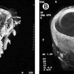
Some patients may be at risk for a cribriform plate fracture, and blindly passing a tube into their nose may result in a nasocerebral (NC) tube (see picture). This is a neurosurgical catastrophe, and the outcome is uniformly dismal. It generally requires craniectomy to remove the tube.

The following patients are at risk:

  • Evidence of midface trauma (eyebrows to zygoma)
  • Evidence of basilar skull fracture (raccoon eyes, Battle’s sign, fluids leaking from ears or nose)
  • Coma (GCS<8)

If you really need the tube, what can you do? If the patient is comatose, it’s easy: just insert an orogastric (OG) tube. However, that is not an option in awake patients; they will continuously gag on the tube. In that case, lubricate a curved nasal trumpet and gently insert it into the nose. The curve will safely move it past the cribriform plate area. Then lubricate a smaller gastric tube and pass it through the trumpet.

http://video.ted.com/assets/player/swf/EmbedPlayer.swf

Printing A Whole Kidney?

And so it begins. I wrote recently about using a special version of a 3D printer to print a skin graft (read it here). I also speculated that we could be seeing 3D printers that could eventually print entire organs at some point. Well, leave it to Wake Forest again.

They demonstrated the concept of printing an organ at the 2011 TED Conference (Technology, Entertainment, Design) last week in Long Beach. Now, this was not a working organ, just a concept demo of sample tissue. Nonetheless, this is a preview of things to come. Imagine when we can print up a new kidney to replace the shattered one in the pan on the back table of the OR.

Check out this fascinating video!

Dry Seal Chest Tube Suction Systems

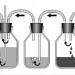
The original chest tube collection system traditionally consisted of three chambers. The picture above shows the classic three bottle system (which I actually remember using during residency). On the left is the suction control bottle that determines how much suction is applied to the patient. The middle bottle provides one way flow of air out of the patient, the so-called water seal bottle. Finally, the right bottle collects any fluid from the pleural space.

Collection systems used in hospitals are much more tidy than this, wrapping all three into one modular unit. However, if you look closely you can identify parts of the system that correspond to each of the bottles.

The problem with the older systems is that they typically require water in the “water-seal” chamber to maintain one-way flow out of the patient. If this chamber is compromised by knocking the system over (see this post), air may be able to enter the patient’s chest, giving them a big pneumothorax.

Management of chest tube collection systems by EMS is tough. It’s very easy to tip the system during air or ground ambulance runs, putting the patient at risk. Some manufacturers have developed so called “dry-seal” systems that use a mechanical one-way valve to avoid this problem.

I have not been able to use one of these systems yet. Here is my take on the pros and cons:

  • Pro – immune to tip-over and malfunction of the water-seal chamber
  • Con – more difficult to detect an air leak. Current models require either injection of a small quantity of water, tipping the system, or converting to a water-seal system.
  • Con – no literature regarding safety of this relatively new technology

Bottom line: Looks like a great idea to me, especially for EMS use. Once they get to the hospital, the unit can be changed to a water-seal system or a larger dry-seal system with the water injection port inthe dry-seal chamber.

Enoxaparin And Pregnancy

Lovenox

Pregnant women get seriously injured, too. And pregnancy is an independent risk factor for deep venous thrombosis. We reflexively start at-risk patients on prophylactic agents for DVT, the most common being enoxaparin. But is it safe to give enoxaparin during pregnancy?

Studies have looked at drug levels in cord blood when the mother is receiving enoxaparin, and none has been found. No specific bleeding complications have been identified, either. So from the baby’s standpoint, administration is probably safe.

However, there are two other issues to consider. In a study looking at the use of enoxaparin for prophylaxis in women with a mechanical heart valve, 2 of 8 women (and their babies) died. Both suffered from clots that developed and blocked the valves. Most likely, the standard dose of enoxaparin was insufficient, so monitoring of anti-Factor Xa levels must be done.

The other problem lies in the multi-dose vial of Lovenox (Sanofi-Aventis). Each 100mg vial contains 45mg of benzyl alcohol, which has been associated with a fatal “gasping syndrome” in premature infants. The individual dose syringes do not have this preservative.

Bottom line: It is probably safe to give enoxaparin to pregnant women after trauma. However, it is unclear if the dose needs to be increased to achieve adequate prophylaxis. Only consider using this medication after consultation with the patient’s obstetrician, and use only the individual dose syringes. Otherwise fall back to standard subcutaneous non-fractionated heparin (even though it is a Category C drug by FDA; it is still considered the anticoagulant of choice during pregnancy).