Tag Archives: blunt trauma

Practical Tip: Evaluation of Hematuria in Blunt Trauma

Bloody urine is a relatively uncommon finding in blunt trauma patients. Hematuria ranges from microscopic to gross. Microscopic means blood that can only be seen with a microscope, and gross means visible to the naked eye. In trauma, we only care about gross hematuria, which ranges from the faintest of pink to the deepest red.

In the picture above gross hematuria is present in all tubes but the far right one. Those four will need further evaluation.

In trauma, gross hematuria is a result of an injury to kidney, ureter or bladder. Blunt injury to the ureter is so rare it’s reportable, so you can pretty much forget that one unless the mechanism is extreme. So you really just need to focus on kidney and bladder.

Any victim of blunt trauma that presents with visible hematuria needs to be evaluated by CT of the abdomen and pelvis with an added CT cystogram. Standard CT technique is done without a urinary catheter, or with the catheter clamped. This is not acceptable for hematuria evaluation, as only 50% of bladder injuries show up with this technique.

CT cystogram is an add-on to the standard CT, and consists of the administration of contrast into the bladder which is then kept under pressure while the scan is performed. Delayed slices through the pelvis after the bladder is depressurized and emptied is routine. Nearly 100% of bladder injuries are detected using this technique.

If the CT shows a renal laceration or hematoma, the patient should be admitted and managed according to your solid organ injury protocol. Kidney injuries fare better that livers and spleens, and only rarely require surgery. If no kidney or bladder injury is seen, the default diagnosis of a renal contusion is the culprit. No treatment is needed, and the patient can be discharged if no other injuries are present. The blood will clear over a few days, but may disappear and reappear a few times in the process. Be sure to warn the patient that this may occur, or you may receive some surprise phone calls. The patient can followup with their primary care physician in a week or two.

The majority of these injuries do not require urologic consultation. Complex injuries with extravasation of urine out of the kidney, or injuries to the collecting system should be referred to a urologist, however.

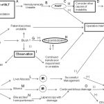
Algorithm For Nonoperative Management of Blunt Hepatic Trauma

Yesterday, I posted the Western Trauma Association’s algorithm for operative management of blunt liver trauma. Click here to view it. Today, I’m going to discuss their algorithm for nonoperative management. 

The algorithm is fairly self-explanatory. Click on the image above to read the annotated text for details on each step. Note: this requires full access to the Journal of Trauma.

Some key points in this algorithm:

  • Unstable patients need rapid identification of the cause. If the FAST is positive ©, then you need to go to the OR and use the operative algorithm.
  • CT scan is used for diagnosis in stable patients (F), but if a liver injury is seen and they become unstable at any time, go to the OR.
  • Contrast extravasation in a stable patient should prompt an evaluation and possible embolization by interventional radiography (G).
  • If complications develop (SIRS, abdominal pain, fever, jaundice), a repeat CT is indicated (K).
  • Abscesses and focal collections of bile may be managed by interventional radiology (L,M). Persistent bile leak may be decreased by ERCP and sphincterotomy (O).
  • Bile ascites or large hemoperitoneum may be managed using laparoscopy with drainage (N).

Reference: Western Trauma Association critical decisions in trauma: nonoperative management of adult blunt hepatic trauma. J Trauma. 67:1144–1148, 2009.

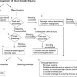
Algorithm For Operative Management of Blunt Hepatic Trauma

The Western Trauma Association has just published guidelines on decision-making when faced with hepatic injury in the OR. The algorithm is based on the available literature, which contains little prospective, randomized trial data. Nonetheless, it is a valuable tool that can be used to develop your own institution-specific protocol.

The algorithm is fairly self-explanatory. Click on the image above to read the annotated text for details on each step. Note: this requires full access to the Journal of Trauma.

Some key points in this algorithm:

  • Simple hemostatic maneuvers are usually successful with minor bleeding (A).
  • Sequential use of more involved maneuvers is indicated for major bleeding. In order, they are packing (B), Pringle maneuver (D), selective vessel ligation within the liver (E), and finally selective hepatic artery ligation (F).
  • Damage control laparotomy and interventional radiology are useful adjuncts.

Tomorrow I’ll write about the nonoperative blunt hepatic trauma algorithm. Click here to view it.

Reference: Western Trauma Association/Critical Decisions in Trauma: operative management of adult blunt hepatic trauma. J Trauma 71(1):1-5, 2011.

Carotid and Vertebral Artery Injury From Blunt Trauma

Blunt injury to the carotid or vertebral arteries (BCVI) is relatively uncommon, but potentially very deadly. Up to 2% of patients with high energy blunt trauma suffer this injury. Many are not diagnosed until the patient has ischemic symptoms or a stroke. However, more aggressive screening has shown a higher incidence that previously thought and may allow intervention before neurologic injury occurs.

Recently, a series of 222 patients with 263 BCVI was retrospectively reviewed, with an eye toward effectiveness of interventions. A total of 29 strokes occurred in the hospital in these patients, but only 7 of these occurred after diagnosis of the BCVI. Mortality was much higher in the stroke group (34% vs 7%). The authors looked at both medical and interventional therapies.

This paper identified the following items:

  • Car crash was the most common mechanism of injury (81%)
  • Vertebral arterial injury was slightly more common than carotid artery BUT
  • Women were much more likely to sustain a carotid injury
  • Older patients were more likely to have a vertebral injury

These authors found that CT angio was not sufficiently sensitive to identify all BCVI. They recommend a formal 4-vessel arteriogram in patients with a negative CT angio who have significant risk factors (unexplained neurologic deficit, Horner’s syndrome, LeFort II or III injury, cervical spine injury, soft tissue injury of the neck).

If a BCVI is identified, the patient should be heparinized until all other injuries are definitively managed. At that point, they should be preloaded with clopidogrel and aspirin and a repeat arteriogram should be performed. Endovascular stenting using a bare metal stent should be performed when possible because it results in the lowest stroke rate and requires the shortest duration of anti-platelet therapies. Patients then continue on aspirin and clopidogrel for an appropriate period of time.

To download the algorithm used by the authors, click here.

Reference: Optimal outcomes for patients with blunt cerebrovascular injury (BCVI): tailoring treatment to the lesion. J Am Coll Surg 212(4):549-559, 2011.

Less Morbidity From Negative Trauma Laparotomies?

Trauma surgeons generally dread the negative laparotomy for trauma. Previous work has shown that complications occur in anywhere from 22% to 53% of cases. Those studies were usually retrospective and included patients with penetrating trauma, which may have skewed the results.

A newly published study tries to throw this common wisdom in doubt. It was a retrospective review of a prospectively maintained database of trauma admissions after blunt trauma . Patients were separated into groups who underwent immediate, delayed or no laparotomy, as well as whether they had or did not have associated injuries. Complications were tracked using an accurate and validated tracking system. The complications tracked included death, DVT, PE, infections, pulmonary issues, as well as other organ system problems.

The authors found that a negative laparotomy did not increase the complication rate, but that a delayed laparotomy did. They also noted that a Complication Impact Score (that they made up) was higher in the delay to laparotomy patients. So they believe that when clinical and imaging findings are equivocal, doing an operation to establish a diagnosis is justifiable.

My Bottom Line: This study does not look at really delayed complications like small bowel obstruction, which we see with some regularity in old trauma patients. Also, other studies have also shown that brief observation, even in patients with a bowel injury, does not increase complications significantly. Unless the potential injury that you are observing is known to have significant complications, my practice is to observe equivocal cases in order to avoid more complications down the road.

Reference: “Never be wrong”: the morbidity of negative and delayed laparotomies after blunt trauma. J Trauma 69(6): 1386-1392, 2010.