Tag Archives: vertebral artery

Diagnosing BCVI In Children

Several days ago, in my post on “How Common Is BCVI?” I mentioned a paper recognizing the increasing incidence of BCVI in pediatric patients and the very high stroke rate (37%) and death rate (13%). These numbers are very concerning!

Previous work shows that the Memphis and Denver criteria are not very sensitive in adults. This has led many trauma centers to add CT angiography of the neck automatically in patients with a high-energy mechanism. But what about kids? Are these screening criteria any better?

A consortium of children’s hospital trauma centers has a paper currently in press that evaluated both the Memphis and Denver criteria in children under age 15. It was a four-year prospective, observational study of children with head, face, or neck injuries. Although the Memphis criteria were specifically used in the study, data for applying the original and expanded Denver criteria, EAST practice management guideline criteria, Utah score, and McGovern score were also collected. The last two are pediatric-specific criteria.

Any child who met at least one of the Memphis criteria received a CTA or MRA of the head and neck. In addition, all children with head, face, or neck injuries received a follow-up evaluation two weeks after discharge. This was designed to capture any evidence of BCVI in those who did not meet Memphis criteria and hence had no CTA/MRA. Patients who missed this evaluation or had other missing data were excluded from the analysis.

Here are the factoids:

  • A total of 2,284 children met the criteria for enrollment; nearly one-third were excluded due to no imaging/follow-up or missing data (!)
  • There were 24 BCVI diagnosed (1.6%)
  • Diagnostic accuracy of the various screening criteria were:
Criteria Sens Spec PPV NPV # CTA to detect one BCVI
Memphis 92 71 5 100 20
Denver 73 88 9 100 11
Expanded Denver 88 64 4 100 25
EAST 79 83 7 100 14
Utah 49 96 16 99 7
McGovern 75 90 11 100 9

The Memphis criteria had the highest sensitivity and would have missed the fewest BCVI. The pediatric-specific Utah score had the highest specificity but would have missed more than half of the injuries. The authors recommend refining the Memphis criteria to improve its specificity while maintaining its high sensitivity.

Bottom line: As with adults, we struggle with systematically identifying BCVI. All screening systems leave something to be desired. It’s not practical or prudent to treat children the same as adults and just liberalize the use of CTA. Substituting MRA is not practical because this requires sedation and/or intubation in the younger age groups.

Of interest in this study, the overall incidence was higher (1.6% here vs. less than 0.5% in my previous post). This is probably due to the fact that there was a significant effort to identify criteria for angiography, and follow-up was provided to detect occult injuries.

This paper adds to the previous work I cited describing how important it is to detect this injury. The current research demonstrates that the Memphis criteria are the best we have for pediatric patients at this time. But it clearly shows the need for a better tool. 

But until one is developed, a best practice would be to use the Memphis criteria to screen any pediatric patient with head, neck, or facial trauma due to a high-energy blunt mechanism. Then select CTA or MRA after conferring with your pediatrics and radiology teams.

Reference: Diagnostic accuracy of screening tools for pediatric blunt cerebrovascular injury: An ATOMAC multicenter study. Journal of Trauma, publish ahead of print, DOI: 10.1097/TA.0000000000003888.

 

Treatment Of BCVI

In my last post, I reviewed the grading system for blunt carotid and vertebral artery injury (BCVI). Today, we’ll discuss treatment, and in the next post, we will wrap up with pediatric-specific information.

There are basically three modalities at our disposal for managing BCVIantithrombotic medication (heparin and/or antiplatelet agents), surgery, and endovascular procedures. The choice of therapy is usually based on surgical accessibility and patient safety for anticoagulation. We know that several studies have shown decreased stroke events in heparinized patients. Unfortunately, this is not always possible due to associated injuries. Antiplatelet agents are usually tolerated after acute trauma, especially low-dose aspirin. However, several studies have shown little difference in outcomes in patients receiving heparin vs. aspirin/clopidogrel for BCVI.

So what to do? Here are some broad guidelines:

  • Grade I (intimal flap). Heparin or antiplatelet agents should be given. If heparin can be safely administered, it may be preferable in patients needing other surgical procedures since it can be rapidly reversed by stopping the infusion. These lesions generally heal entirely on their own, so a follow-up CT angiogram should be scheduled in 1-2 weeks. Medication can be stopped when the lesion heals.
  • Grade II (flap/dissection/hematoma). These injuries are more likely to progress, so heparin is preferred if it can be safely given. Stenting should be considered, especially if the lesion progresses. Long-term anti-platelet medication may be required.
  • Grade III (pseudoaneurysm). Initial heparin therapy is preferred unless contraindicated. Stable pseudoaneurysms should be followed with CTA every six months. If the lesion enlarges, surgical repair should be performed in accessible injuries or stenting in inaccessible ones.
  • Grade IV (occlusion). Heparin therapy should be initiated unless contraindicated. Patients who do not suffer a catastrophic stroke may do well with follow-up antithrombotic therapy. Endovascular treatment does not appear to be helpful.
  • Grade V (transection with extravasation). This lesion is frequently fatal, and the bleeding must be addressed using the best available technique. For lesions that are surgically accessible, the patient should undergo the appropriate vascular procedure. Inaccessible injuries should undergo angiographic treatment and may require embolization to control bleeding without regard for the possibility of stroke.

References:

  1. Scott WW, Sharp S, Figueroa SA, et al. Clinical and radiographic outcomes following traumatic Grade 1 and 2 carotid artery injuries: a 10-year retrospective analysis from a Level I trauma center. J Neurosurg 122:1196, 2015.
  2. Scott WW, Sharp S, Figueroa SA, et al. Clinical and radiographic outcomes following traumatic Grade 3 and 4 carotid artery injuries: a 10-year retrospective analysis from a Level 1 trauma center. J Neurosurg 122:610, 2015.
  3. Scott WW, Sharp S, Figueroa SA, et al. Clinical and radiological outcomes following traumatic Grade 1 and 2 vertebral artery injuries: a 10-year retrospective analysis from a Level 1 trauma center. J Neurosurg 121:450, 2015.
  4. Scott WW, Sharp S, Figueroa SA, et al. Clinical and radiological outcomes following traumatic Grade 3 and 4 vertebral artery injuries: a 10-year retrospective analysis from a Level I trauma center. The Parkland Carotid and Vertebral Artery Injury Survey. J Neurosurg 122:1202, 2015.

How To Grade BCVI

In my last post, I reviewed the three screening systems for blunt carotid and vertebral artery injury (BCVI). Today, we’ll look at grading them.

Just ten years ago, it was a major production to identify BCVI. Then, CT angiography was still in its early days, and scanner resolution and radiologist experience were significant issues that impacted accuracy.

We’ve come a long way in a relatively short period of time, and current-day scanners now have more than adequate resolution. It’s also more common to have a radiologist with special skills reading these studies, the neuroradiologist. For these reasons, CT angiography has become the standard for diagnosis. It is also the most cost-effective. Only in sporadic cases do we need to obtain a conventional contrast angiogram.

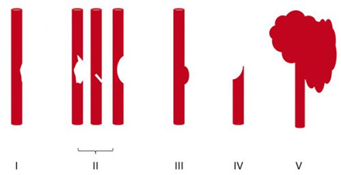
Once the study has been obtained, it’s time to identify and classify the injury. The Denver group is also responsible for bringing us the grading system for BCVI. See the diagram below.

Here are the details:

Grade I: A mild intimal irregularity is seen. Note the abnormally narrowed area, representing a minor intimal injury, possibly with a small amount of clot.

Grade II: This grade has several presentations. There may be an intraluminal thrombosis/hematoma with (left) or without (right) an intimal flap, or a flap alone (center).

Grade III: There is a full-thickness injury to the vessel with a contained extraluminal extravasation (pseudoaneurysm)

Grade IV: The vessel is completely occluded by a flap or thrombus

Grade V: The artery is transected and freely extravasating

Here’s a nice diagram:

Remember, we always grade things for a reason! Ultimately, the injury grade will translate into the selection of treatment. We’ll cover that in my next post.

Reference: Blunt carotid arterial injuries: implications of a new grading scale. J Trauma. 1999;47(5): 845-53.

How Common Is BCVI?

Blunt carotid and vertebral artery injuries (BCVI) are an under-appreciated problem after blunt trauma. Several screening tools have been published over the years, but they tend to be unevenly applied at individual trauma centers. I will discuss them in detail in the next section.

For the longest time, the overall incidence of BCVI was thought to be low, on the order of 1-2%. This is the number I learned years ago, and it has not really changed over time.

But how do we know for sure? Well, the group at Birmingham retrospectively reviewed every CT angiogram (CTA) of the neck they did in a recent two-year period. They did this after adopting a policy of imaging each and every one of their major blunt trauma patients for BCVI. Each patient chart was also evaluated to see if the patient met any of the criteria for the three commonly used screening systems.

During the study period, a total of 6,287 of 6,800 blunt trauma patients underwent BCVI screening with CTA of the neck. They discovered that 480 patients (7.6%) were positive for BCVI!

This is a shocking 8x higher than we expected! So why hasn’t this been obvious until now? Most likely because we were previously only aware of patients who became symptomatic. Luckily, many of these patients dodge the proverbial bullet and never exhibit any symptoms at all.

And what about pediatric patients? The neurosurgery groups at the University of New Mexico and Texas Children’s Hospital analyzed data in the Kids’ Inpatient Database (KID), which contains nationally representative pediatric data from the US. Five samples were obtained three years apart, beginning in 2000 and extending to 2012.

There were nearly 650,000 admissions for blunt trauma in the database, and 2150 were associated with BCVI. There was an interesting trend: incidence in 2000 started at 0.24% and increased to 0.49% in 2012. This represents a relative doubling of cases! Keep in mind that the absolute numbers remained very small, especially compared to the adult incidence.

Children aged 4 to 13 had the lowest risk of sustaining BCVI. This was higher in younger kids (ages 0-3), probably due to their big heads. It was also higher in adolescents and young adults (age 15-20). The injury was found more often in conjunction with cervical spine, skull base, clavicle, and facial fractures, as well as in children with TBI and intracranial hemorrhage.

Over one-third of children sustaining BCVI suffered a stroke (37%). Mortality was high, with a total mortality of 13%. This increased to a 20% rate if a stroke occurred.

So why should we be worried? This is one of those clinical entities like blunt thoracic aortic disruption that potentially has terrible consequences if ignored. And it seems to be worse among children even though it is far less common. Although the number of patients who develop sequelae from their BCVI is small, suffering a stroke can be catastrophic.

Should we perform a screening study for all blunt trauma patients? It seems like overkill, or is it? Is there any way we can be more selective about it?

In the next post, I’ll review the current screening tools used to determine which patients should receive CTA and how good they are.

References:

  1. Universal screening for blunt cerebrovascular injury. J Trauma 90(2):224-231, 2021.
  2. Blunt cerebrovascular injury in pediatric trauma: a national database study. J Neurosurg Pediatr. 2019

Blunt Carotid And Vertebral Artery Injury (BCVI): First Adults, And Now In Children!

I’ve written quite a bit about the challenges of diagnosing blunt carotid and vertebral artery injury (BCVI) in adults.  And now some papers on the potential danger to pediatric patients are beginning to surface.

I think it’s time to repost and update my series on BCVI. I’ll start with the basics, like nomenclature. I’ll then move on to how often we actually encounter it. This will include new information on younger patients.

I’ll dig into the various screening systems, and will include new information from some recent pediatric papers. The, I’ll finish up with how to grade it and suggested treatment routines based on grade.

Lets start with the acronym itself. There seems to be some confusion as to what BCVI actually stands for. Some people believe that it means blunt cerebrovascular injury. This is not correct, because that term refers to injury to just about any vessel inside the skull.

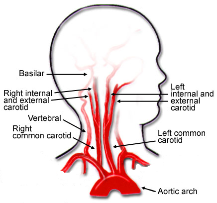
The correct interpretation is blunt carotid and vertebral artery injury. This term refers to any portion and any combination of injury to those two pairs of vessels, from where they arise on the great vessels, all the way up into the base of the skull. Here’s a nice diagram:

Note that we will be excluding the external carotid arteries from this discussion, since injuries to them do not have any impact on the brain. They can cause troublesome bleeding, though.

These arteries are relatively protected from harm during blunt trauma. But given enough energy, bad things can happen. Fortunately, injuries to these structures are not very common, but unfortunately many trauma professionals under-appreciate their frequency and severity.

In the next post, we’ll explore the incidence of this injury in both adults and children. Is it truly as uncommon as we think?