Tag Archives: Tracheostomy

Tracheostomy After Anterior Cervical Fusion: Can It Be Too Soon?

Early tracheostomy is generally accepted to be a good thing in critically injured patients. A number of papers have shown that it decreases ventilator days and hence ICU and hospital days,  and also reduces pneumonia rates, sedation requirements, and saves quite a bit of money.

However, there has been one problematic group in whom critical care surgeons are often forbidden to do an early trach: patients with a recent anterior cervical fusion. These patients typically have unstable cervical fractures, with or without a concomitant spinal cord injury. The spine surgeons argue that doing a trach “too soon” leads to a higher infectious complication rate due to the proximity of the trach to their anterior surgical wound.

But is this really true? The trauma/critical care program at Thomas Jefferson University in Philadelphia looked at their experience. They are a federally sponsored spinal cord injury center, and have a vast experience compared to most trauma centers. They reviewed their experience over a 16 year period. Typically, they performed all of their tracheostomies within 10 days, so they arbitrarily defined “early” as within 4 days, and “late” as > 5 days after the cervical procedure.

Here are the factoids:

  • A total of 98 patients with tracheostomy after anterior fusion were included in the study, some of whom also underwent a concomitant posterior fusion
  • 39 cases were “early”, within 4 days of the anterior fusion procedure, and 59 were “late”
  • Average time to fusion in the early group was 2 days, and 10 days in the late group
  • There were no wound infections in the early group
  • There were 5 wound infections in the late group, and 4 of them involved the posterior fusion site(!)
  • The only infection of the anterior fusion site occurred in a late patient who suffered an esophageal perforation from the fusion hardware

Bottom line: Although the numbers are still small after 15 years of data, it’s probably the best we will ever get! It is clear that an anterior fusion wound is safe in these procedures. I am at a loss as to why the posterior fusion wounds tend to get infected, though. But the next time your spine surgeons balk about doing an early trach in one of their anterior fusion, show them this paper!

Percutaneous Tracheostomy Without The Bronchoscope

It’s always nice to find an article that supports your biases. I’ve been doing percutaneous tracheostomy since the 1990’s, and have used a variety of kits and equipment over the years. Some of these turned out to be rather barbaric, but the technique is now quite refined.

A routine part of the procedure involved passing a bronchoscope during the procedure to ensure that the initial needle was placed at the proper level and in the tracheal midline. It was also rather frightening to watch the trachea collapse when the dilators were inserted.

I abandoned using the bronchoscope in this procedure about 15 years ago. It was an annoyance to get the bronchoscope cart and a respiratory therapist to help run it. And to find someone available to pass the scope while I did the trach. So I added a little extra dissection to the technique, directly visualizing the trachea at the desired location. From then on, I had no need to see the puncture from the inside because I could see it quite well from the outside!

An article in the Journal of Trauma demonstrated that this technique works just as well without the scope. The authors looked at their own series of 243 procedures; 32% were done with the bronchoscope, 68% without. There were 16 complications overall, and the distribution between the bronch and no-bronch groups was equal.

Bottom line: In general, the bronchoscope is not needed in most percutaneous tracheostomy procedures. It adds complexity and expense. However, there are select cases where it can be helpful. Consider using it in patients in a Halo cervical immobilizer, the obese, or in patients with known difficult airway anatomy. And always do the more difficult ones in the OR, not the ICU.

Reference: Percutaneous tracheostomy: to bronch or not to bronch – that is the question. J Trauma 71(6):1553-1556, 2011.

In my last post, I discussed the Passy-Muir valve, which allows patients who have a tracheostomy tube in place, and are not on a ventilator, to talk. But what about patients who are still vent dependent? It’s very frustrating for both patient and trauma professionals when we can’t communicate with each other.

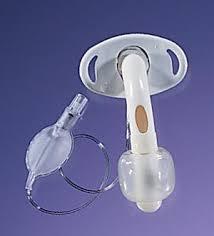
Pulmodyne, Inc. makes the Blom tracheostomy tube system, which solves this problem. This device has a large fenestration in the back of the tube with a special bubble valve (see below), coupled with an inner cannula that has a 1-way flap valve. This allows controlled release of air into the pharynx, enabling speech while on the ventilator.

image

A multicenter study looked at voice production and intelligibility of speech in a group of 23 ventilated patients with a trach tube in place. Although not entirely clear in the paper, it appears that all were changed to Blom trach tubes for the study (2 had one in place at the beginning of the study). Overall, voice production and intelligibility were good. Most were able to begin audible speech within about 6 minutes of initial application. One deconditioned patient took longer. The video below shows an example of the speech that is achievable.

Bottom line: This novel product allows a subset of trauma patients to speak while still on the ventilator. It is most appropriate for those who do not have significant head injury, especially those with facial trauma requiring airway protection with a tracheostomy.

Related post:

I have no financial interest in Pulmodyne, Inc.

What Is: A Passy-Muir Valve?

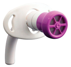
Some critically injured patients undergo tracheostomy due to prolonged ventilatory failure. As they recover, the trach tube is usually downsized over time until it can be permanently removed. Unfortunately, this process may take a month or more, and the patient is generally unable to speak during this time. Writing and other forms of communication are both slow and frustrating, so a Passy-Muir valve may be attached to the trach to allow (nearly) normal speech during the recovery process.

image

The Passy-Muir is a one way valve that allows air to be inhaled through the trach tube, but not exhaled through it. Instead, the air must circulate around the tube and up through the trachea to the pharynx. This particular brand has features that help keep the tube from collecting secretions during speech. See the brief video from the manufacturer below.

The downside to this device is that it increases the work of breathing during exhalation because air must flow around the entire trach tube, which is narrowing the available tracheal lumen. Note: if the trach tube has a balloon, it must be deflated or the patient will not be able to breathe! Some patients do not have enough strength to overcome this narrowing initially, and may not be able to speak or say more than a few words at a time. This improves with practice, and helps speed up tube removal.

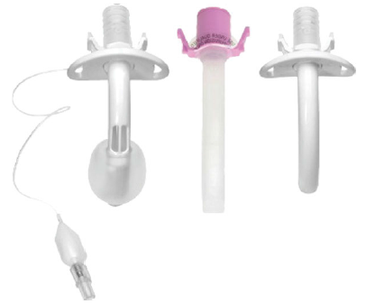
Use of a fenestrated tracheostomy tube with the Passy-Muir is helpful, because it has a strategically placed (but small) hole that allows air flow through a portion of the tube. When this type of tube is used, the balloon must also be deflated, since it is subject to mucus plugging which would completely obstruct the airway. Work of breathing is still increased because this hole is small relative to the diameter of the trachea.

image

Note: I have no financial interest in Passy-Muir Inc or any other tracheostomy product manufacturers.

Percutaneous Tracheostomy Without The Bronchoscope

It’s always nice to find an article that supports your biases. I’ve been doing percutaneous tracheostomy since the 1990’s, and have used a variety of kits and equipment. Some of these turned out to be rather barbaric, but the technique is now quite refined. 

A routine part of the procedure involved passing a bronchoscope during the procedure to ensure that the initial needle was placed at the proper level and in the tracheal midline. It was also rather frightening to watch the trachea collapse when the dilators were inserted.

I abandoned using the bronchoscope in this procedure about 10 years ago. It was an annoyance to get the bronchoscope cart and a respiratory therapist to help run it. And to find someone available to pass the scope while I did the trach. So I added a little extra dissection to the technique, directly visualizing the trachea at the desired location. From then on, I had no need to see the puncture from the inside because I could see it quite well from the outside!

An article in the Journal of Trauma shows that this technique works just as well without the scope. The authors looked at their own series of 243 procedures; 32% were done with the bronchoscope, 68% without. There were 16 complications overall, and the distribution between the bronch and no-bronch groups was equal.

Bottom line: In general, the bronchoscope is not needed in most percutaneous tracheostomy procedures. It adds complexity and expense. However, there are select cases where it can be helpful. Consider using it in patients in a Halo cervical immobilizer, the obese, or in patients with known difficult airway anatomy. And always do the more difficult ones in the OR, not the ICU.

Reference: Percutaneous tracheostomy: to bronch or not to bronch – that is the question. J Trauma 71(6):1553-1556, 2011.