Tag Archives: penetrating injury

The Role Of Postop CT Scan In Penetrating Trauma

CT scans are commonly used to aid the workup of patients with blunt trauma. They are occasionally useful in penetrating trauma, specifically when penetration into a body cavity is uncertain, and the patient has no hard signs that would send him or her immediately to the operating room.

Is there any role for CT in operative penetrating trauma, after the patient has already been to the OR? The dogma has always been that the eyeballs of the surgeon in the OR are better than any other imaging modality. Really? The surgical group at San Francisco General addressed this question by retrospectively reviewing 6 years of their operative penetrating injury registry data. They were interested in finding how many occult injuries (seen with CT but not by the surgeon) were found on a postop CT. A total of 225 patients who underwent operative management of penetrating abdomen or chest injury were included. Here are the factoids:

  • Only 110 patients had a postop CT scan; 73 had scans within the first 24 hours, the other 37 were scanned later
  • The rationale for early scan was to investigate retroperitoneal injury in half of patients, but frequently no indication was given (41%)
  • The rationale for late scan was for workup of ileus in one-third or for evaluation of new or unexpected clinical problems
  • Occult injuries were found in about half of early CT patients (52%) and 22% of late CT patients
  • The most common occult injuries were fractures, GU issues, regraded solid organ injury, and unrecognized vascular injuries
  • Ten patients had management changes, including:
    • Interventional radiology for four injuries with extravasation
    • Operation for orthopedic or GU injury in seven patients
    • One patient underwent surgery for an unstable spine fracture

Bottom line: There appears to be a significant benefit to sending some penetrating injury patients to CT in the early postop period. Specifically, those with injury to the retroperitoneum, deep into the liver, near the spine, or with multiple and complicated injuries would benefit. Simple stabs and gunshots that stay away from these areas/structures probably do not need follow-up imaging. 

Reference: Routine computed tomography after recent operative exploration for penetrating trauma: What injuries do we miss? J Trauma 83(4):575-578, 2017.

The Evolution Of Penetrating Neck Trauma Management – Part 3: Determining Risk

In the last post, I described the first crucial step in the contemporary management of penetrating neck trauma, control of obvious external hemorrhage. Let’s move on to the nuts and bolts of figuring out what needs to be done about the injury.

Now, it’s time to triage your patient based on clinical signs that predict the presence or absence of a significant injury. In the old days, the neck was conceptualized as three different zones that dictated the diagnostic and management algorithm.

We are now moving toward considering the neck as a single unit. The next decision point is to determine the risk for vascular or aerodigestive tract injury based on an examination for signs of injury. These signs have been divided into three groups.

Hard signs. These indicate a high risk for deeper injury and consist of the following:

  • Vascular signs
    • Refractory shock
    • Pulsatile or difficult-to-control hemorrhage
    • Large or expanding hematoma
    • Audible bruit or palpable thrill (I hardly ever see anyone actually check the neck for these, so brush up your skills!)
  • Aerodigestive signs
    • Airway compromise or stridor
    • Bubbles from the wound
    • Significant subcutaneous emphysema
    • Major hematemesis
    • Massive hemoptysis
  • Neurologic signs
    • Neurologic deficits that suggest embolic strokes from a vascular injury

Soft signs. These suggest an intermediate risk for injury and are:

  • Vascular signs
    • Small or stable hematoma
    • History of bleeding or hypotension that has resolved
    • Active venous oozing
    • Pulse volume or blood pressure discrepancy (this suggests a thoracic vascular injury)
  • Aerodigestive signs
    • Hoarseness or any voice changes
    • Painful swallowing
    • Difficult swallowing
    • Mild subcutaneous emphysema
    • Minor hematemesis
    • Minor hemoptysis
  • Neurologic signs
    • Local neurologic deficit (direct injury to local nervous structures)

No signs. Obviously, this suggests a low risk of injury.

Once the level of risk has been determined, a course of action can be planned. Most patients with hard signs will require operative intervention. Plain x-rays with skin markers in place may help visualize retained foreign bodies and their relationship to bony structures. If the signs are immediately life-threatening, this step should be skipped, and operative exploration should be performed immediately. If the patient is stable and the injuries may be outside the easily accessible area of the neck (the old Zone II), a multi-detector CT angiogram (MDCTA) may help with operative planning. It may also identify patients eligible for endovascular repair.

Patients with soft signs have a lower risk of injury and should immediately undergo MDCTA. This scan has very high sensitivity and specificity in this group.

Finally, patients with no signs of deeper injury rarely need any intervention. Small series suggest that these patients could potentially be discharged from the ED. However, most trauma professionals will be uncomfortable with the thought of this. MDCTA is a low-risk test; until we know better, it’s probably best to obtain it before discharge.

Bottom line: I have described the initial assessment and management of patients with penetrating neck injury using the newer method using signs of injury in place of the old zones of injury. Nuances are still possible, such as what to do if the MDCTA is indeterminate for a vascular or aerodigestive injury. Fortunately, that is fodder for another post!

Reference: Approach to Penetrating Neck Trauma: What You Need to Know. J Trauma Acute Care Surg. 2024 Mar 25. doi: 10.1097/TA.0000000000004292. Epub ahead of print. PMID: 38523116.

The Evolution Of Penetrating Neck Trauma Management – Part 2: Initial Steps

In my previous post, I described the early days of penetrating neck injury management and introduced a paper suggesting that this concept should be revised. Today, I will summarize a paper by Siletz and Inaba that is currently in press and outlines what the contemporary way of treating these injuries should be.

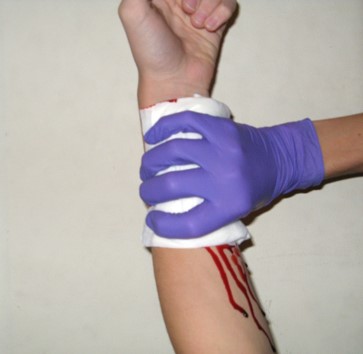
Step 1. If present, rapidly control external hemorrhage and airway compromise. As always, bleeding should be controlled by direct pressure or packing. Direct pressure does not look like this:

The goal is to create a zone of pressure higher than the systolic BP perfectly in the area of bleeding. Since pressure is force per unit area, a larger area like that show above diffuses the maximum pressure and just doesn’t work. Note the ongoing bleeding shown in the picture.

Here’s what direct pressure looks like:

Or

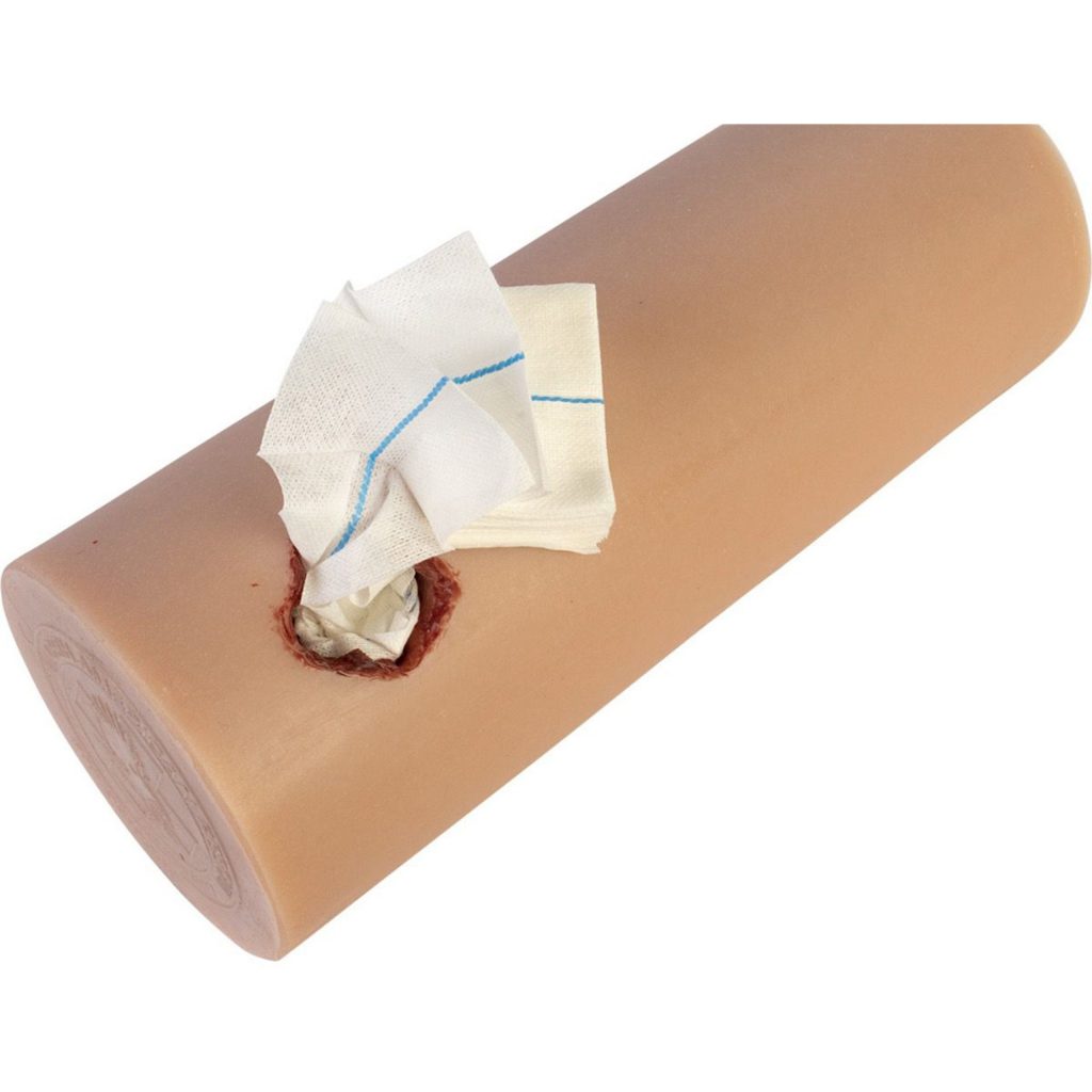
A single finger (or maybe two) should be placed on or in the wound. If deeper bleeding is a problem, the same kind of pressure can be accomplished by packing with gauze. If gauze is used, however, pressure must usually be applied over the gauze to make sure that the underlying tissues remain pressurized.

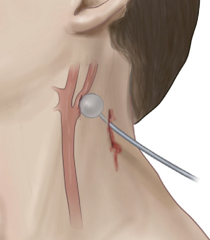
If gauze packing is not practical because of this need for additional pressure, a urinary catheter can be inserted into the wound and inflated until the bleeding stops.

Courtesy Core EM

Airway control should ideally occur in the operating room. Given the proximity of this wound to airway structures, it is imperative that an ideal environment is present when the airway is inserted. A skilled anesthesiologist should be present, with difficult airway equipment available if needed. The surgeon should be standing by with all equipment needed to obtain a surgical airway if needed. Even though the patient may be breathing okay, the airway structures may be distorted by hematoma or injury.

You have probably noted that this is the same initial assessment we used in the old three zones approach. In the next post, I will discuss the details of a new assessment approach that considers the neck a single unit.


The Evolution Of Penetrating Neck Trauma Management – Part 1

“When the facts change, I change my mind. What do you do, sir?”

This is a famous quote from John Maynard Keynes. (Or is it? There is some debate over its authenticity, but you get the idea it tries to convey.) Our knowledge base continually changes, so we must be willing to change our minds (and practices) based on new, reliable information.

The management of penetrating neck injury is one of those facets in trauma care that has undergone slow but steady progress over the past 40 years of my career. In the old days, we quickly identified the zone of injury and proceeded to the operating room for Zone II injuries. We had to think a little harder about the other zones to be certain that we needed to be in the OR. But overall, the threshold for surgery was low.

Things have been changing. Five years ago, I published a post detailing new work by Inaba et al. at LAC+USC. This started a move toward using more straightforward criteria and advanced imaging to assist decision-making with these injuries.

In this post, I’ll summarize the original paper. In the next section, I will describe the group’s paper, which is currently in press and outlines the full framework for workup or penetrating neck injury.

The advance that makes this new method possible is based on the high degree of accuracy that CT angiography of the neck provides. It is very sensitive for identifying even minor injuries to the aerodigestive tract and vascular system.

The trauma group at LAC+USC organized a prospective, multicenter study using a multidetector CT angiography of the neck for initial screening of penetrating neck injury. This allows the evaluation of the neck as a single unit, not as three zones. It also solves the problem of trying to apply zones to injuries that cross several of them.

The new algorithm that was tested utilized an initial physical exam, first looking specifically for “hard signs” of injury.  The following were considered the hard signs:

  • Active hemorrhage
  • Expanding or pulsatile hematoma
  • Bruit or thrill over the injured area
  • Unresponsive shock
  • Hemoptysis or hematemesis
  • Air bubbling from the wound

These patients were immediately taken to the OR and explored through an appropriate incision.

Patients with no signs or symptoms were admitted and observed for at least 24 hours. All other patients were considered to have “soft signs.” They underwent multidetector CT angiography of the neck, with a scanner having at least 40 slices. Further evaluation of these patients was based on the exam and CT scan.

Here are the factoids:

  • 453 patients with penetrating neck injuries were identified during the 31-month study period
  • 9% had hard signs and were taken to the OR; 50% had soft signs and underwent CT; 41% had no signs and were observed
  • For soft sign patients, 86% of scans were negative, and all were true negatives after observation
  • 12% of soft sign patients had a positive scan, and of those, 81% were true positives
  • four patients (2%) with soft signs had too much artifact for an accurate CT, and other tests were performed; 1 of the 4 had an injury
  • Sensitivity of CTA was 100%, and specificity was 97.5% in the soft sign patients
  • The authors concluded that CTA is very reliable for identifying injuries in patients with soft signs and that patients with no signs do not require scanning, only observation

Bottom line: This was an intriguing paper that utilized both physical examination and CT angiography. The results were impressive, and they supported the argument that CTA is not required in all stable patients. With additional numbers and time, it has become clear that we can safely adopt this algorithm. My next post will flesh out the details.

Reference: Evaluation of multidetector computed tomography for
penetrating neck injury: A prospective multicenter study. J Trauma 72(3):576-584, 2012.

Use Of Radio-opaque Markers In Penetrating Trauma

As I was browsing through my journal list this week, I ran into an interesting title for an article that is currently in press.

“The use of radio-opaque markers is medical dogma”

Catchy, especially since I love writing about dogma vs what is really supported by the literature. The author questions the justification of this practice and posits that there are risks to extrapolating information based on radiographs with markers placed by the trauma team.

OLYMPUS DIGITAL CAMERA

The author first reviewed the literature on the use of markers for penetrating injury, which started only recently, in 2002. Markers were initially used to precisely locate the penetration site since skin wounds (obviously) don’t show up on X-rays. Typically, these were just plain old paper clips. Some trauma professionals placed them directly over the wound. Others un-bent them and fashioned them into shapes that pointed to the exact location of the wound.

With the growing usage of CT scans to evaluate stable patients, modifications to the marker were made. Small arrow markers designed for use on x-rays were frequently used. However, even the very small ones could cause enough scatter on a CT scan to interfere with diagnosis. At some centers, Vitamin E capsules were taped on top of the wound. But thankfully, there are now special markers that can pinpoint the wound without degrading the tomographic image.

The author goes on to describe how gunshot wounds specifically are difficult to assess with a marker. Although the exact surface location may be noted, the underlying injuries vary due to size, distance, velocity, and trajectory change from tissue density or bone strikes. He also notes that it may not be wise to place a marker into a bloody field in a potentially combative patients.

The article concludes that the use of this technique for identifying anything other than surface location of penetrations lacks clinical evidence and is based only on expert opinion. Which essentially makes it dogma.

Bottom line: Here are my thoughts. First, the use of markers on penetrating wounds has been going on for much longer than the 20 years found in the trauma literature reviewed here. It has been a common practice among trauma surgeons for many, many decades. Most “seasoned” (old) trauma surgeons have been doing and teaching this for their entire careers. 

I concur that we have techniques like CT scan available to us now that provide an excellent view of the penetration trajectory. The skin wound is usually apparent on the scan, but may be improved with the use of a CT-approved marker.

So why still do this for the patient arriving in your trauma bay? An experienced trauma surgeon can get a good sense of the trajectory based on the entry point, the exit wound, and the location of any retained bullet or fragments. Rapid placement of some kind of marker on all wounds followed by a quick image allows them to roughly predict what was hit, and assess the possibility that there might be bleeding that would drive the team straight to the operating room. It can help direct the surgical exploration if imaging was unnecessary or contraindicated. 

So yes, this is dogma. The reality is that no one will ever be able to design a study that definitively evaluates the very soft outcomes that result from using this technique. But every senior trauma surgeon can easily cite numerous examples in their career when this method has been extremely useful. The lack of a study only means that there will never be any evidence-based guideline for the use of this technique. However, it is still acceptable to have a protocol based on substantial clinical experience. But as with all dogma, once that definitive study finally does comes along, the protocol must be modified to adhere to the findings of the study.

For now, keep using those markers! And I’m very interested in comments from both old and young trauma professionals on this topic.

Reference: The Use of Radio-opaque Markers is Medical Dogma, doi:10.1111/acem.1485, Dec 2023.