Tag Archives: fracture

Yes, Smoking is Bad!

Everybody knows that smoking is bad. But how often have you stopped by to see one of your trauma patients and have been told “they’re out smoking?” Well, it turns out it’s bad for their injuries as well.

A German group looked at the effects of smoking on healing of a “simple” tibial fracture. They looked at 103 patients who underwent treatment for an isolated tibial shaft fracture at a trauma center. Patients with more complicated problems like extension into a joint, open fracture (Gustilo III), or significant soft tissue injury were excluded. 

Patients were divided into non-smokers and smokers (including previous smokers). A total of 85 patients were studied, and there were roughly half in each group. The nonsmoking group experienced no delayed or non-unions of their fractures. The smoking group reported 9 delayed unions and 9 non-unions in 46 patients! As expected, time off work and eventual functional outcome was worse as well.

Bottom line: The exact mechanism for impairment of fracture healing by smoking is unclear. It may be due to physiologic effects of inhaled tobacco components on blood flow, blood vessels, transforming growth factor levels or collagen formation. It could also be a secondary effect of socioeconomic variables, patient compliance, or a host of other factors. Regardless, it’s bad. Smoking should be forbidden while in hospital, and should be strongly discouraged after discharge.

Reference: Cigarette smoking influences the clinical and occupational outcome of patients with tibial shaft fractures. Injury 42:1435-1442, 2011.

Fracture Blisters Demystified

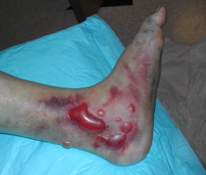
Fracture blisters pop up (!) in trauma patients now and then, and nobody seems to know what to do with them. Here’s a primer on dealing with them.

A fracture blister typically occurs near fractures where the skin has little subcutaneous tissue between it and bone. These include elbows, knees, ankles and wrists. They tend to complicate fracture management because they interfere with splinting, casting, and incision planning for open reduction procedures. They can appear anytime within a few hours of injury to 2-3 weeks later.

These blisters are thought to be caused by shearing forces applied at the time of injury. There are two types described, based on their color: clear fluid and hemorrhagic. The difference lies in the level of the shear. Clear fluid blisters have separated within the epidermis, and hemorrhagic blisters separate at the dermal-epidermal junction. The clinical difference is healing time; clear blisters take about 12 days and hemorrhagic blisters heal in about 16 days. 

So should we pop the blisters and operate/splint, or wait for them to heal and then go to surgery? Unfortunately, there’s no great data on this and it usually hinges on the preferences of the orthopaedic surgeon. Waiting delays care an average of 7 days, and longer for tibial plateau and calcaneal fractures. Operating immediately anecdotally increases wound infection rates.

Bottom line: Anticipate fracture blisters by looking at location and severity of mechanism. Try to schedule operative reduction as soon as is practical. And monitor the wound closely to make sure that delayed blisters don’t cause complications due to splinting or casting.

Pelvic Binder Orthosis vs Pelvic External Fixation

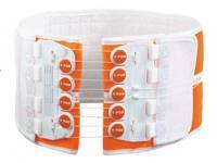
In the “old” days, the recommended management for an unstable pelvis was application of an external fixator. In some textbooks, it was even suggested that this should be done (by orthopedics) in the resuscitation room. High volume trauma centers with ortho residents could actually pull this off, but not many others.

As the idea of pelvic orthotic binders caught on (T-POD, sheeting, etc) and was adopted by prehospital providers, and then trauma teams, the use of initial external fixation dropped off. But the idea that external fixation was the most desirable or most effective lingered on. A study from Memphis finally sheds some light on the answer to this question.

A 10 year retrospective review was carried out on patients presenting with multiple or severe pelvic ring fractures who had early stabilization of the pelvis. Stabilization consisted of external fixation early on, and gradually shifted to pelvic orthotic devices over the study period. They ultimately analyzed outcomes for 93 patients in each treatment group.

The authors found that transfusion needs were dramatically reduced with the orthotic devices (5 units vs 17 units at 24 hours) compared to the orthotics. About a quarter of patients in each group went to angiography, and even in those patients the transfusion need remained lower in the orthotic device group. Hospital length of stay was also significantly shorter in this group (17 vs 24 days). There was no difference in mortality.

Bottom line: Although this is a small, retrospective study it easily showed significant results and will probably never be repeated. Use of a pelvic orthotic device (POD) resulted in less blood replacement and shorter stays in hospital. This technique is simple, cheap and quick, an ideal combination. But does a sheet count as an orthotic device? We don’t know. It’s really cheap, but probably a bit less controlled than a POD. If you have a real POD in your ED or your ambulance, use it. If not, apply the sheet as described below.

Reference: Emergent pelvic fixation in patients with exsanguinating pelvic fractures. JACS 204:935-942, 2007.

New Technology: Fracture Putty

Fracture healing takes a long time, as many of our patients can attest to. Six or eight weeks, and even more may be required for full healing. Researchers at the University of Georgia and in Houston have completed an animal study on rats using a type of “fracture putty” that dramatically speeds up this process. 

The researchers used adult mesenchymal stem cells that produce a protein which is involved in bone healing and regeneration. They created a gel using these cells, and injected them into the fracture sites which were stabilized externally (imagine a rat external fixator!). The fractures healed rapidly, and within 2 weeks the rats could run and stand on their legs normally.

Bottom line: The next step is to translate this work to larger animals. Strength and durability are major concerns. The amount of stress placed on rat legs and human legs is considerably different. If this pans out, it could revolutionize fracture healing, especially in cases where there may be highly disabling segmental bone loss (read: military). It will be several years before this can move to human studies.

Reference: University of Georgia Regenerative Bioscience Center

Temporal Bone Fracture

The temporal bone is a very complicated and important piece of bone located at the base of the skull. It houses a number of structures vital to hearing and balance, and is a conduit for several important nerves. Most fractures are due to blunt trauma, and although several classification systems exist, the majority are irregularly shaped and don’t lend themselves to easy categorization.

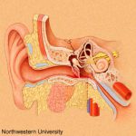
Clinically, a t-bone fracture should be suspected anytime that blood is seen in the ear canal or behind the eardrum after blunt head trauma. Patients may complain of dizziness or hearing loss. Examination may demonstrate some nystagmus in some cases. Most of the time, the diagnosis will not be made until a head CT is performed. The diagram above shows why this fracture can cause hearing problems (middle ear) or dizziness (inner ear).

There is usually no specific surgical intervention needed for this fracture. However, two important functional exams must be completed once diagnosed: facial nerve function and hearing. The facial nerve exam should document whether only portions of the nerve are affected vs all branches, and whether there is at least partial function. In head injured patients that can’t follow commands, even a grimace can demonstrate some function.

The ideal hearing test is carried out with a tuning fork. However, this is not usually convenient in acute trauma patients. Whispering and rubbing fingers near the patient’s ear can be used as a quick and dirty test. Awake patients will be able to tell you if their hearing is decreased.

Bottom line: Suspect temporal bone injuries in patients with any signs of basilar skull fracture. If the diagnosis is suspected or confirmed by CT scan, document the best facial nerve and hearing exam that you can. Contact your facial surgeon once the diagnosis is confirmed, and call them urgently if there is any loss of facial nerve function. Thin cut CT scans of the temporal bone are generally not necessary, and should not be ordered automatically unless the facial surgeon needs it for a specific reason.