Tag Archives: fat embolism

Diagnosis Of Fat Embolism Syndrome

A number of scoring systems have been developed to identify FES (Gurd’s and Wilson’s criteria, Schonfeld’s criteria, Lindeque’s criteria to name a few). Unfortunately, none of these are helpful. They were developed in the 1980s as part of the authors’ studies on the use of  steroids for treatment, and no one else has taken the time to study their sensitivity and specificity.

Diagnosis of FES is primarily clinical. It relies upon recognition of the principal findings on physical exam, and exclusion of more common conditions that may mimic it.

Here is a template for diagnosing FES:

Is your patient at risk? The vast majority of these patients will have fractures. One, or especially two or more long bone fractures (mostly the femur) are usually present. Other fractures that add risk are those involving the pelvis or bones that contain marrow, such as the ribs and sternum. Patients who have just undergone fracture repair are also at risk and will be discussed in the next section. Finally, patients who have had intraosseous lines placed are also at risk, regardless of the type of infusate.

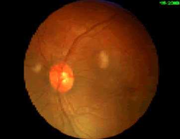
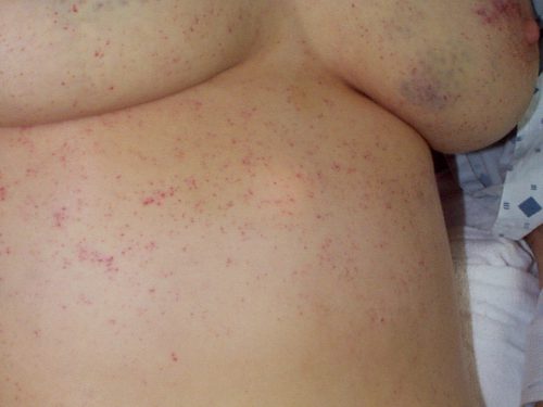
What signs or symptoms have developed? Skin changes are very suggestive of FES if your patient is at risk. However, rashes are common manifestations of contact allergies, drug reactions, infectious diseases, and many other conditions. If those are ruled out, then the presence of risk factors plus a rash is sufficient to make the diagnosis.

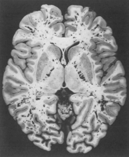
Mental status changes are more difficult to pin on FES, even though it is a more common initial presentation than the rash. Since this is a trauma patient, you must rule out delayed manifestations of head trauma. Urgent CT of the head is required to do so. And typically, there will be no specific findings that point to FES. It is always a diagnosis of exclusion.

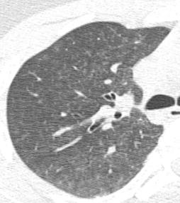
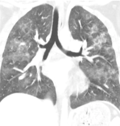
Pulmonary dysfunction requires a search for the usual suspects. A good physical examination of the chest coupled with a chest x-ray will help identify pneumothorax, hemothorax, or pneumonia. A chest CT may be indicated if pulmonary embolism is suspected.

Once other more common clinical problems have been eliminated, you are left with the diagnosis of FES. There are no specific lab tests to draw, and more invasive studies are neither helpful nor indicated. Fat embolism syndrome is a diagnosis of exclusion.

Next, the relationship of fat embolism and orthopedic surgery.

Clinical Manifestations Of Fat Embolism Syndrome

There are three organ systems that are classically involved in FES: pulmonary, CNS, and skin. Manifestations generally begin between 24 and 72 hours after injury. In rare cases, symptoms can begin within 12 hours. In my experience, these tend to be the ones that become the most severe and are frequently life-threatening.

Pulmonary (95% of cases): This is the most common manifestation of FES, and may occur without other signs and symptoms. Nearly all patients develop some degree of hypoxia. Progressive tachypnea and mild tachycardia may provide the first clinical clue if oxygen saturation is not being monitored.

Chest x-ray is usually unremarkable early on. And once the syndrome has developed, it is generally not helpful. CT scan is useful for defining the extent of pulmonary injury, but lags the clinical picture by several days. Findings are non-specific, usually consisting of small, ground-glass opacities in the periphery.

In the example above, the opacities are very small and difficult to see.

But they’re a little more obvious here!

Other CT findings include small pulmonary nodules in the upper lobes or along peripheral pulmonary vessels. These are thought to be areas of obstruction caused by the emboli. Nonspecific pleural effusions may be seen, and bronchial thickening has also been described. Rarely, fat globules may be seen in the lower extremity veins or IVC, and should immediately raise suspicion for developing FES even before symptoms develop.

CNS (60% of cases): If they occur, CNS changes generally crop up after the pulmonary manifestations begin. Generally, they start as mild confusion, but can progress to decreasing level of consciousness and even coma. Focal neurologic deficits are occasionally seen, and seizures can occur.

The actual mechanism behind this appears to be very similar to the skin changes which will be described in the next section. Emboli occur in vessels predominantly in the white matter of the brain. This leads to petechial hemorrhages, which are likely due to the inflammatory mechanisms previously described.

Note the numerous dark petechiae visible in the white matter in this specimen.

Retinal exam can also show evidence of fat embolism. Fat globules may actually be seen in the retinal vessels early.

Note the fat globules at the 9:30 and 2:00 positions to the optic nerve in the image above.

Skin (33% of cases): The most recognizable sign of FES is the petechial skin rash. This rash usually involves the torso, and axillary petechiae are very common. It can spread to involve the head and neck, and occasionally the extremities. Subconjunctival hemorrhages are sometimes seen. The rash tends to be transient and usually lasts only a few days. Here is an example of the classic petechial rash.

Other findings: Fat globules may be found in the urine in patients with FES. However, they are commonly present in patients with long bone fractures, so their presence is not helpful or predictive. Nonspecific findings such as fever, leukocytosis, anemia, and thrombocytosis are also relatively common. In severe cases, cardiac dysfunction, hypotension, and peripheral hypoperfusion can occur. I have personally seen necrosis of fingers and toes from a very severe case.

Unfortunately, the “classic” triad of mental status changes, skin rash, and pulmonary insufficiency are seen in only a small minority of patients. Typically, only one or two signs and symptoms appear at the same time, making diagnosis a bit challenging.

Tomorrow, making the diagnosis of fat embolism syndrome.

Fat Embolism vs Fat Embolism Syndrome

It’s fat embolism week! Fat embolism syndrome (FES) is one of those clinical problems that trauma professionals read about during their training, then rarely ever see. Although the clinical manifestations are frequently mild, they can progress rapidly and become life-threatening. Over the next five days, I’ll try to  help you better understand this condition, and provide details on diagnosis and treatment.

Fat embolism syndrome (FES) is a constellation of findings that arise from a single, unified cause: the escape of fat globules into the circulation (fat embolism). The ultimate resting places of those globules determine the specific manifestations of FES seen in clinical practice. When it occurs, it typically becomes apparent 24 to 72 hours after injury.

Simple fat embolism occurs to some degree any time tissues containing fat are manipulated or injured. It has been demonstrated during plastic surgical injections for cosmetic purposes and lipid infusions. It is more frequently seen with orthopedic injuries, especially those involving the femurs and pelvis. And it makes sense that the more fractures that are present, the more likely fat embolism will occur. Embolism is also known to occur when performing orthopedic procedures, particularly those involving the marrow cavity (intramedullary nailing), but has also been reported in total knee and hip procedures.

Fat embolism syndrome has a generally reported incidence of 1 – 10%, although I believe that is on the high side. I see a case every 3 – 4 years in a predominantly blunt, fracture-laden practice. Fat embolism without symptoms occurs much more frequently. A study from 1995 using transesophageal echo found evidence of emboli in 90% of patients with long bone fractures.

But how do these fat globules get into the circulation and produce such chaos? We know that they can be mechanically pushed into small venules when tissues containing fat cells or bone marrow are injured. In bone, there are numerous small venules located throughout that are anchored to it. When the bone is fractured, these venules tear and are held open so yellow (fatty) marrow can be pushed into them.

If enough emboli enter the blood stream, they may accumulate in the end vessels of tissues and block flow. Although this is a simple and appealing explanation, it may not be the full story. If the emboli primarily occur during and after injury, why does it take several days for the full-blown syndrome to develop?

A likely explanation is that the fat globules begin to degrade while in the circulatory system. Breakdown into free fatty acids results in the release of a cascade of cytokines and other mediators. The inflammatory response around the end vessels create the gross pathology that we associate with fat embolism syndrome.

Tomorrow, clinical manifestations of fat embolism syndrome.

The February 2019 Trauma MedEd Newsletter Is Here!

Welcome to the current newsletter. This issue is devoted to an uncommon yet somewhat troubling problem, fat embolism. Here are some of the things I cover:

  • Fat Embolism vs Fat Embolism Syndrome (FES)
  • Clinical Manifestations Of FES
  • Diagnosis Of Fat Embolism Syndrome
  • FES And Orthopedic Surgery
  • Prevention And Treatment Of Fat Embolism Syndrome

The March issue will be released to subscribers late this month and will cover a potpourri of interesting little tidbits. I’ll release it to everyone via this blog in April, so subscribe now if you want it sooner!

To download the current issue, just click here! Or copy this link into your browser: http://bit.ly/TME201902.