Tag Archives: embolization

Complications of Splenic Embolization for Trauma

Angioembolization has become a common procedure that can increase the likelihood of success for nonoperative management for splenic trauma. It does have its own set of complications to be aware of, however.

The most obvious complication is mechanical injury to the femoral artery. This occurs in 1 to 3% of patients. It is more common in the very young (small caliber artery) and the elderly (arteries of stone). Rarely, the substance or device that is used for the embolization may migrate or end up on the wrong spot, infarcting something important.

A common issue that occurs is infarction of portions of the spleen. This is actually the desired effect, as it stops the bleeding. Most of the time, we are unaware of the changes that take place in the spleen post-procedure. But every once in a while we get a repeat CT scan days or weeks down the road and see some very interesting things.

The most common finding is a splenic infarct alone. This is an area of the spleen, sometimes wedge shaped, that does not take up contrast. This is normal. In some cases, gas bubbles are seen within the spleen parenchyma, usually within the infarcted area. In others, large areas of gas are present, and an air-fluid level may also be seen. This is definitely not normal.
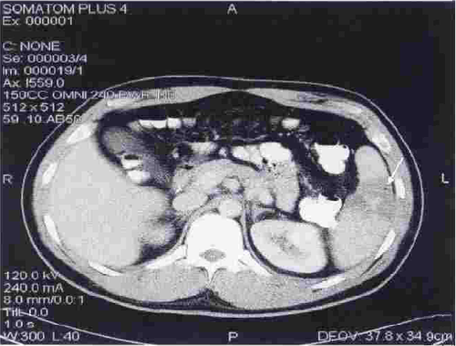
Note the infarcted area at the arrow, with a tiny gas bubble visible.

Tiny bubbles are normal after this procedure, and can be ignored if the patient does not appear ill and does not have any systemic evidence of inflammation or sepsis. On the other hand, big bubbles or air-fluid levels probably indicate a developing splenic abscess, and the patient will usually appear ill and have a high WBC count. Unfortunately, the only treatment for this is splenectomy. Insertion of drainage catheters does not work and the patient will only become sicker if it is attempted.

Post-Embolization Syndrome?

Important! If your search for a trauma topic brought you here…

The Trauma Professional’s Blog has moved to this new site, but old Google or Bing searches may link you to this page.

Please retype your search in the box in the upper left corner of any page (or touch the magnifying glass above on a mobile device) to find what you are looking for.

Go to theTrauma Professional's Blog page

And hopefully, all the search engines will finish indexing the new site soon! Explore the site and enjoy! Michael

———————————————————————————-

I’ve seen a number of trauma patients who have developed pain and elevated WBC after embolization of solid organs for trauma. For kidneys and main splenic artery embolization, it’s fairly common in my experience. Turns out, this phenomenon was described in 2007-2008 in patients undergoing embolization of hepatic tumors and uterine fibroids. It was termed post-embolization syndrome, and consists of pain, fever, nausea and ileus.

An article was just published in the Journal of Trauma describing this syndrome in children after splenic embolization for blunt trauma. The authors looked at their own trauma registry over a 12 year period. Yes, it took that long to find 448 children with blunt splenic injury. Of those, only 11 underwent arterial embolization (sigh of relief).

The average age was about 13 and ISS was 16 in both groups. Kids who underwent embolization were more likely to spend some time in the ICU, had a longer hospital stay (8 vs 5 days(!)), and took longer to resume their diet (5 vs 2 days). These differences occurred despite the fact that most of the embolized children had isolated splenic injuries. Additionally, the embolized children were more likely to receive blood (3 units vs none) and plasma.

My first question about this paper is, why? Broken spleens in children do not act like broken spleens in adults. The vast majority of the cases of contrast extravasation in children stops on its own without intervention. So why did we even have to find out that post-embolization syndrome occurs in children? They shouldn’t be going through this procedure anyway! Fortunately, a deeper read of the paper provides the answer. The indication for angio was splenic pseudoaneurysm in 2, and ongoing hemorrhage in the other 9. In the case of these latter 9, it did keep the children from having their spleens operated on.

Bottom line: In general, don’t send kids for splenic angiography (99.3% of kids in this study did not have it). Ongoing hemorrhage (prior to hypotension, which is an absolute indication for OR) is probably the only indication I can think of. Pseudoaneurysm and extravasation of contrast are not indications like they are in adults. But if you do have to send them, just be aware that they may develop pain, fever and ileus that will keep them in the hospital and/or ICU for a few extra days.

Reference: Transarterial embolization in children with blunt splenic injury results in postembolization syndrome: A matched case-control study. J Trauma 73(6):1558-1563, 2012.

Spleen Embolization After Trauma

Angioembolization of the spleen (AES) is part of our armementarium in the management of spleen and liver trauma. However, there are no good guidelines to help us decide exactly which patients would benefit from it. An abstract to be presented at the EAST meeting in January 2013 gives us a little more information on the actual benefits of this procedure.

The authors did a retrospective review of the management of blunt splenic injury at four busy Level I trauma centers. They looked at 1275 injured patients over a 3 year period. Here are the interesting tidbits from the study:

  • There was considerable variation in the use of AES at the 4 centers, ranging from 1% of patients to 14%. This should be no surprise because there is no real guidance available yet.
  • There was also a large degree of variation between the number of initial splenectomy performed at these centers
  • Centers that used AES more frequently had lower initial splenectomy rates
  • Patients at centers with high AES rates were 3 times more likely to leave with their spleen intact

Bottom line: This abstract correlates with my own personal experience: judicious use of angioembolization saves spleens. The real question is about which patients are best served by it. Our protocol is to strongly consider it in all high grade spleen injuries (Grade 4 and 5), and to always do it if a blush or extravasation is present. Our success rate for nonoperative management currently stands at about 94%.

Related posts:

Reference: Variation in splenic artery embolization a spleen salvage: a multicenter analysis. EAST Annual Scientific Assembly, Paper #1, to be presented January 2013.

Angioembolization For Splenic Injury

Initial nonoperative management of splenic injury is standard in hemodynamically stable patients. Over the past decade, the success rates have climbed by adding angioembolization to the algorithm, according to several published series. However, the objective benefit and specific indications have not been worked out.

A paper published this month by the University of Florida, Jacksonville used the NTRACS registry to try to clarify these issues. They identified 1039 patients undergoing nonoperative management (NOM) over a nearly 10 year period. Patients who died shortly after arrival, those who went directly to OR for hemodynamic reasons, and children were excluded, leaving 539 patients. Only about 1/6 of the patients underwent embolization. 

The overall failure rate was about 4%, a little higher in the non-angio patients, a little lower with angio. Incidentally, the angio group had significantly higher injury severity (26 vs 20). Analysis of the lower grade spleen injury group showed no improvement in success rate by adding angio. However, the high grade groups (grades IV-V) did benefit by adding this procedure. Similarly, success improved when performing angio in patients with contrast blush or evidence of slow, ongoing bleeding. If NOM did fail, it usually occurred on day 2.

Bottom line: Although we’ve been adding angio to non-operative management of spleen and liver injury for a decade, here’s the first paper that has been able to define the real indications for doing it. First, all unstable patients go to the OR (don’t even consider nonop management). In the remaining patients, if the CT shows a grade IV or V injury, or a contrast blush, angio is recommended. If neither of these is noted, but the hemoglobin continues to decline “too quickly” (surgeon judgement), then a trip to angio is also warranted. Applying these principles can increase your success rate to about 96%.

Related post:

Reference: Selective angiographic embolization of blunt splenic traumatic injuries in adults decreases failure rate of nonoperative management. J Trauma 72(5):1127-1134, 2012.

Complications of Splenic Embolization for Trauma

Angioembolization has become a common procedure that can increase the likelihood of success for nonoperative management for splenic trauma. It does have its own set of complications to be aware of, however.

The most obvious complication is mechanical injury to the femoral artery. This occurs in 1 to 3% of patients. It is more common in the very young (small caliber artery) and the elderly (arteries of stone). Rarely, the substance or device that is used for the embolization may migrate or end up on the wrong spot, infarcting something important.

A common issue that occurs is infarction of portions of the spleen. This is actually the desired effect, as it stops the bleeding. Most of the time, we are unaware of the changes that take place in the spleen post-procedure. But every once in a while we get a repeat CT scan days or weeks down the road and see some very interesting things.

The most common finding is a splenic infarct alone. This is an area of the spleen, sometimes wedge shaped, that does not take up contrast. This is normal. In some cases, gas bubbles are seen within the spleen parenchyma, usually within the infarcted area. In others, large areas of gas are present, and an air-fluid level may also be seen. This is definitely not normal.

Tiny bubbles are normal after this procedure, and can be ignored if the patient does not appear ill and does not have any systemic evidence of inflammation or sepsis. On the other hand, big bubbles or air-fluid levels probably indicate a developing splenic abscess, and the patient will usually appear ill and have a high WBC count. Unfortunately, the only treatment for this is splenectomy. Insertion of drainage catheters does not work and the patient will only become sicker if it is attempted.