Category Archives: General

What A MESS! Part 1

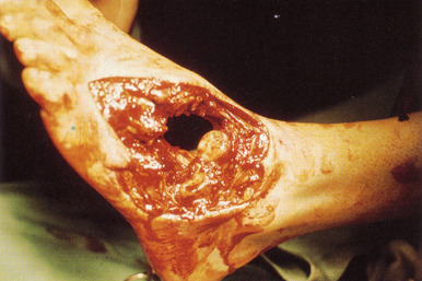
The Mangled Extremity Severity Score (MESS) is now 25 years old, and it still serves us fairly well. This simple system helps predict salvageability of mangled extremities. Obviously, the acronym was chosen to help describe the clinical problem.

The system was originated at the Harborview Medical Center in Seattle. The development was not very scientific; the authors put their heads together and made a list of the four things that they observed predicted limb salvage:

  • Degree of skeletal and soft tissue injury
  • Presence of limb ischemia
  • Presence of shock
  • Age

The system was used retrospectively in a group of 25 patients(!) and the authors found a nice breakpoint at 7. Any mangled extremities with a MESS of 7 or more required amputation. They then applied this to 26 patients prospectively(!) and got the same result.

As you can see, the numbers were small, and there was no followup information. Nevertheless, MESS still stands today, and the critical MESS score has not changed much. It has been validated by a number of other studies during the past 20 years. It is conceivable that the critical score will slowly creep upward with advancements in flap coverage and surgical technique, but it hasn’t done so yet.

Tomorrow, I’ll show you how to calculate the MESS score, and give some tips on how to use it.

Reference: Objective Criteria Accurately Predict Amputation Following Lower Extremity Trauma. Johansen, et al. J Trauma 30(5): 568, 1990.

What The Heck? Final Answer

So what is going on with your dyspneic patient after partial ejection from their car? They are not responding to your interventions as they should How is he immune to chest tube placement?

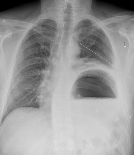
Well, you could intubate the patient. But the x-ray will be available momentarily, and may guide you along a different path. It might even obviate the need for intubation. So while you are getting your meds ready (just in case), this image comes up:

The chest tube is in good position, but the stomach is not!

The reason your patient didn’t respond to the chest tube is that they have a traumatic rupture of the diaphragm with the stomach in the chest. It is compressing a good portion of the left lung, leading to dyspnea and poor oxygen saturation. 

Rapidly place a nasogastric tube to try to decompress the stomach. It doesn’t always work because the angles at the hiatus are not what they usually are. But if it does, the patient will feel better immediately. You may be able to avoid intubating them… for a few minutes. This is a surgical problem, and commonly involves injury to other abdominal structures, especially the spleen. Order up some blood, and rapidly complete your evaluation. A pelvis x-ray is in order, because fractures are common after full or partial ejection through the window. No other imaging is necessary. The send your patient off to the OR for a thorough exploration and repair. Now you can intubate.

I’ve seen this injury three times after partial ejection, and always involving the driver (left side, makes sense). Any time you have left sided abnormalities after blunt trauma that don’t respond to a chest tube, think about this problem.

What The Heck? The original presentation

What The Heck? Part 2

Thanks to all who tweeted, commented and emailed their suggestions! The case involves a partially ejected patient who is brought to the ED with respiratory distress and diminished lung sounds on the left. 

Sounds easy, right? But remember, this is the Trauma Professional’s Blog! I want you to be prepared for things that are a little outside the ordinary. No zebras here, but stuff you could actually see.

First, every trauma activation patient gets supplemental oxygen as the festivities continue. You need to quickly figure out if this is an airway, breathing, or circulation problem. Yes, circulation. Major torso vascular injuries and tamponade can cause respiratory distress. However, we would not expect the blood pressure to be anywhere near normal.

So check the airway to make sure there is no foreign material there. Check the trachea for position. This is one of those classic test results in medicine: if the trachea is deviated, they most likely have a tension pneumothorax. But if it’s not, that doesn’t necessarily mean they don’t. In this case, the trachea is in its usual place, but don’t count tension physiology out yet.

Double check the breath sounds. You confirm that they are nearly absent on the left. What to do next?

You must presume some major problem on the left: large hemo- or pneumothorax, or a tension pneumothorax. Since your patient is physiologically abnormal, you cannot wait to get a chest x-ray. You have to deal with the breathing problem right away. The correct answer is to needle the left chest, then follow immediately with a chest tube.

You do so, and both procedures go smoothly. The chest tube fogs with exhalation, and there is a small amount of blood (100cc) that drains into the collection system. But your patient does not look or feel any better! Oxygen saturations are still in the low 80′s, and he remains dyspneic. As you were finishing the chest tube, the radiology tech snapped a quick chest x-ray, and the result will be up in two minutes.

Now what? Your choices are:

  • Intubate
  • Insert another chest tube
  • Package the patient and run to the OR
  • Wait for the chest x-ray

Again, tweet, comment or email. What is wrong, and why didn’t the chest tube work? What is the ideal next move? Answer tomorrow.

What The Heck? The original presentation

What The Heck? Pop Quiz

You are working in your local emergency department and are notified of an incoming trauma patient. The victim was involved in a car crash at highway speeds, was not restrained, and was partially ejected from the driver’s side window.

Pre-arrival report from the medics indicate that he has a BP of 146/90, pulse of 130, and a respiratory rate of 36. He is very dyspneic and complains that he can’t breathe. They state that the only abnormality that they found on their exam was some bony crepitus over the left lateral chest.

When he arrives, he is exactly as billed. O2 saturations are 82%, and he is in obvious respiratory distress. Breath sounds are quite diminished on the left.

What are the potential diagnoses?

What do you do next? Here are your choices:

  • Examine the airway
  • Apply supplemental oxygen
  • Intubate
  • Obtain a chest x-ray
  • Decompress the left chest with a needle
  • Insert a chest tube
  • Proceed to the operating room
  • Obtain a CT scan of the torso

Please tweet or leave comments with your suggestions. I’ll review your choices on Monday, and provide some followup information. Final answer on Tuesday!

Managing Penetrating Injuries: Some Practical Tips

Although penetrating injuries are a relatively uncommon mechanism at most trauma centers, they are more likely than not to injure deeper structures. Key decisions need to be made quickly during the initial evaluation in order to provide the best care.

Here are some practical tips:

  • Penetrating injuries to just about anything but the extremities should activate your trauma team.
  • If your patient is hypotensive, they will need to go to the OR. You can certainly start infusing some fluid or blood, but a lot leaked out before they got to you, indicating that the leak needs to be surgically fixed. No exceptions.
  • All hypotensive patients require activation of your massive transfusion protocol and consideration of giving tranexamic acid (TXA).
  • If your patient is normotensive, you have the luxury of evaluating them more thoroughly. But don’t lose your sense of urgency. Assume they are dying until you prove otherwise.
  • Complete your secondary survey. Don’t skimp on the exam and always look at the back. If your patient ends up on an OR table, it may be the only time you get to look at it for quite some time.
  • Get a single x-ray of the affected area, even if you need to go to the OR quickly. This can help plan your operation, and may drive you to explore areas you had not considered.
  • Before shooting the x-ray, mark any and all entry and exit points. This will help to predict the trajectory and any injured structures.
  • Use small markers, but not too small. Most radiology departments have small arrows, which are ideal. Dots are too small and may not show up well on plain images. But be aware that some markers may be too dense for CT, causing artifacts that may obscure pathology.
  • Watch out for your own safety! Somebody was trying to kill your patient, and they may show up at your hospital to try to finish the job. Make sure your ED and inpatient areas take appropriate security precautions.