You’re running a trauma activation, and everything is going great! Primary survey – passed. Resuscitation – lines in, fluid going. You are well into the exam in the secondary survey.
Then it happens. The automated blood pressure cuff shows a pressure of 72/44. But the patient looks so good!
You recycle the cuff. A minute passes and another low pressure is noted, 80/52. You move the cuff to the other arm. Xray comes in to take some pictures. You roll the patient. 76/50. Well, you say, they were lying on the cuff. Recycle it again.
A minute later, the pressure is 56/40, and the patient looks gray and is very confused and diaphoretic. It’s real! But how long as it been real? An easy 5 minutes have passed since the first bad reading.
Bottom line: Sometimes it’s just hard to believe that your patient is hypotensive. They look so good! But don’t be fooled. If you get a single hypotensive reading, STOP! You have 90 seconds to figure out if it’s real, so don’t do anything else but. Check the pulse rate and character with your fingers. Do a MANUAL blood pressure check. It’s fast and accurate. If you have the slightest doubt, ASSUME IT’S REAL. Remember, your patient is bleeding to death until proven otherwise. And it’s your job to prove it. Fast!
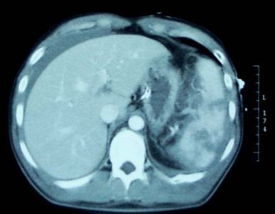
Any trauma professional who has dealt with spleen injuries knows that the white blood cell (WBC) count rises afterwards. And unfortunately, this elevation can be confusing if the patient is at risk for developing inflammatory or infectious processes that might be monitored using the WBC count.
Is there any rhyme or reason to how high WBCs will rise after injury? What about after splenectomy or IR embolization? An abstract is being presented at the Clinical Congress of the American College of Surgeons next month that examines this phenomenon.
This retrospective study looked at a convenience sample of 75 patients, distributed between patients who had splenic injury that was either not treated, removed (splenectomy), or embolized. Data points were accumulated over 45 days.
Here are the factoids:
20 patients underwent splenectomy, 22 were embolized, and 33 were observed and not otherwise treated
Injury severity score was essentially identical in all groups (19)
Splenectomy caused the highest WBC counts at the 30 day mark (17.4K)
Embolized patients had mildly elevated WBC levels (13.1K) that were just above the normal range at 30 days
Observed patients had high normal WBC values (11.0K) after 30 days
Values in observed and embolized patients normalized to about 7K after 30 days; splenectomy patient WBC count remained mildly elevated at 14.1K.
The authors concluded that embolization does not result in permanent loss of splenic function (bad conclusion, rookie mistake!)
Bottom line: This study is interesting because it gives us a glimpse of the time course of leukocytosis in patients with injured spleens. If you need to follow the WBC for other reasons, if gives a little insight into what might be attributable to the spleen. Splenectomy generally results in a chronically elevated WBC count, which tends to vary in the mid-teens range. Embolization (in this study) transiently elevates the WBC count, but it then drops back to normal.
The big problem with this study (besides it being small) is that it fails to recognize that there are many different shades of embolization. Splenic artery? Superselective? Selective? I suspect that the WBC count in main splenic artery embolization may behave much like splenectomy in terms of leukocytosis. And the conclusion about splenic function being related to WBC count was pulled out of a hat. Don’t believe it.
Leukocytosis after Splenic Injury: A Comparison of Splenectomy, Embolization, and Observation. American College of Surgeons Scientific Forum Abstracts pg S164, 2015.
If you need some last minute trauma-related education credits, consider viewing or attending Trauma Education: The Next Generation. It’s tomorrow, beginning at 8:00 am Central Time at Metro State University in St. Paul.
You can watch for free via Livestream. If you want credit, register first and pay a nominal fee. Or join us live in the audience!
Cervical collars are applied to blunt trauma patients all the time. And most of the time, the neck is fine. It’s just those few patients that have fracture or ligamentous injury that really need it.
I’ve previously written about how good some of the various types of immobilization are at limiting movement (click here). But what happens when you are actually putting them on or taking them off? Could there be dangerous amounts of movement then?
Several orthopaedics departments studied this issue using an electromagnetic motion detector on “fresh, lightly embalmed cadavers” (!) to determine how much movement occurred when applying and removing 1- and 2-piece collars. Specifically, they used an Aspen 2-piece collar, and an Ambu 1-piece. They were able to measure flexion/extension, rotation and lateral bending.
Here are the factoids:
There were no significant differences in rotation (2 degrees) and lateral bending (3 degrees) when applying either collar type or removing them (both about 1 degree)
There was a significant difference (of 0.8 degrees) in flexion/extension between the two types (2-piece flexed more). Really? 0.8 degrees?
Movement was similarly small and not significantly different in either collar when removing them
Bottom line: Movement in any plane is less than 3-4 degrees with either a 1-piece or 2-piece collar. This is probably not clinically significant at all. Just look at my related post below, which showed that once your patient is in the rigid collar, they can still flex (8 degrees), rotate (2 degrees) and move laterally (18 degrees) quite a bit! So be careful when using any collar, but don’t worry about doing damage if you use it correctly.
Reference: Motion generated in the unstable cervical spine during the application and removal of cervical immobilization collars. J Trauma 72(6):1609-1613, 2012.
Home of the Trauma Professional's Blog
Do you want to get a daily email every time there’s a new post? See what I’m up to.