Category Archives: General

The Cost Of Duplicate Radiographic Studies

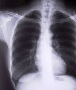
Duplicate radiographic studies are a continuing issue for trauma professionals, particularly after transfer from a smaller hospital to a trauma center. The incidence has been estimated anywhere from 25% to 60% of patients. A lot has been written about the radiation dangers, but what about cost?

A Level II trauma center reviewed their experience with duplicate studies in orthopedic transfer patients retrospectively over a one year period. They looked at the usual demographics, but also included payor, cost information, and reason for repeat imaging. Radiation dose information was also collected.

Here are the factoids:

  • 513 patients were accepted from 36 referring hospitals
  • 48% had at least one study repeated, 256 CT scans and 161 conventional imaging studies
  • Older patients and patients with low GCS were much more likely to receive repeat studies
  • There were no association with the size of the referring hospital or the ability of the patient to pay
  • Most transfers had commercial insurance; only 11% had Medicaid and 17% were uninsured
  • Additional radiation from repeat scans was 8 mSv. The average radiation dose from both hospitals was 38 mSv. This is 13 years of background radiation exposure!
  • The cost of all the repeat studies was over $96,000

Bottom line: This is an eye-opening study, particularly regarding how often repeat imaging is needed, how much additional radiation is delivered, and now, the cost. And remember that these are orthopedic patients, many of whom had isolated bony injuries. I would expect that patients with multiple and multi-system injuries would require more repeat imaging and waste even more money. It is imperative that all centers that receive transfers look at adopting some kind of electronic data transfer for imaging, be it a VPN or some cloud-based service. With the implementation of the Orange Book by the American College of Surgeons, Level I and II centers will receive a deficiency if they do not have some reliable mechanism for this.

“Level I and II facilities must have a mechanism in place to view radiographic imaging from referring hospitals within their catchment area (CD 11–42).”

Reference: Clinical and Economic Impact of Duplicated Radiographic Studies in Trauma Patients Transferred to a Regional Trauma Center. J Ortho Trauma 29(7):e214-e218, 2015.

Why We Can’t Just Stop Anticoagulants In Trauma Patients

We’ve all been faced with injured patients who are taking some kind of anticoagulant, and it complicates their care. Why can’t we just stop them in patients at risk for injury (e.g. an elderly patient who falls frequently)?

Two major risk groups come to mind: those taking the meds who have DVT (or a propensity to get it), and patients with atrial fibrillation who take them to decrease stroke risk. I was not able to find much info (yet) on the former category. But there is a series of nicely done studies based on work from the Framingham Heart Study.

The Framingham study started in 1948, and has been following over 5,000 people for the development of cardiovascular disease. In this particular analysis, 5070 patients who were initially free of disease were analyzed for development of atrial fib and occurrence of stroke. Anticoagulants were seldom used in this group.

The authors found that the prevalence of stroke increased with age in patients with atrial fib. The percentage that could be attributed to a-fib also increased. The following summarizes their numbers:

  • Age 50-59: 0.5 strokes per 100 patients, attributable risk 1.5%
  • Age 60-69: 1.8 strokes per 100 patients, attributable risk 2.8%
  • Age 70-79: 4.8 strokes per 100 patients, attributable risk 9.9%
  • Age 80-89: 8.8 strokes per 100 patients, attributable risk 23.5%

Bottom line: The risk of having a stroke just because a patient has atrial fibrillation goes up significantly with age. So setting an age cutoff for taking an anticoagulant doesn’t make sense. Unfortunately, increasing age also means increasing risk of injury from falls. Warfarin definitely cuts that risk, and it happens to be relatively easily reversbile. However, the newer non-reversible drugs change the equation, shifting the risk/benefit ratio too far toward the dark side. We need some good analyses to see if it really makes sense to move everybody to these new (expensive) drugs just to make it easier to dose and monitor. The existing studies on them only look at stroke, but don’t take injury morbidity and mortality into account.

Reference: Atrial fibrillation as an independent risk factor for stroke: the Framingham study. Stroke 22:983-988, 1991.

Click here to download a reference sheet for dabigatran reversal.

Novel Hip Reduction Technique: The Captain Morgan

I wrote about posterior hip dislocation and how to reduce it using the “standard” technique quite some time ago (see link below). Emergency physicians and orthopedic surgeons at UCSF-Fresno have published their experience with a reduction technique called the Captain Morgan.

Named after the pose of the trademark pirate for Captain Morgan rum, this technique simplifies the task of pulling the hip back into position. One of the disadvantages of the standard technique is that it takes a fair amount of strength (and patient sedation) to reduce the hip. If the physician is small or the patient is big, the technique may fail.

In the Captain Morgan technique, the patient is left in their usual supine position and the pelvis is fixed to the table using a strap (call your OR to find one). The dislocated hip and the knee are both flexed to 90 degrees. The physician places their foot on the table with their knee behind the patient’s knee. Gentle downward force is placed on the patient’s ankle to keep the knee in flexion, and the physician then pushes down with their own foot, raising their calf. Gentle rotation of the patient’s hip while applying this upward traction behind the patient’s knee usually results in reduction.

Some orthopedic surgeons use a similar technique, but apply downward force on the patient’s ankle, using the leverage across their own knee to develop the reduction force needed. The Captain Morgan technique use the upward lift from their own leg to develop the reduction force. This may be gentler on the patient’s knee.

The authors report a series of 13 reductions, and all but one were successful. The failure occurred due to an intra-articular fragment, and that hip had to be reduced in the operating room.

I’m interested in hearing comments from anyone who has used this technique (or the leverage one). And does anyone have any other techniques that have worked for them?

Related post:

Reference: The Captain Morgan technique for the reduction of the dislocated hip. Ann Emerg Med (in press) dol:1016/j.annemergmed.2011.07.010, 2011.

Liver Laceration And Liver Function Tests

Over the years I’ve seen a number of trauma professionals, both surgeons and emergency physicians, order liver transaminases (SGOT, SGPT) and bilirubin in patients with liver laceration. I’ve never been clear on why, so I decided to check it out. As it turns out, this is another one of those “old habits die hard” phenomena.

Liver lacerations, by definition, are disruptions of the liver parenchyma. Liver tissue and bile ducts of various size are both injured. Is it reasonable to expect that liver function tests would be elevated? A review of the literature follows the typical pattern. Old studies with very few patients.

From personal hands-on observations, the liver tissue itself tears easily, but the ducts are a lot tougher. It is fairly common to see small, intact ducts bridging small tears in the substance of the liver. However, larger injuries can certainly disrupt major ducts, leading to major problems. But I’ve never seen obstructive problems develop from this injury.

A number of papers (very small, retrospective series) have shown that transaminases can rise with liver laceration. However, they do not rise reliably enough to be a good predictor of either having an injury, or the degree of injury. Similarly, bilirubin can be elevated, but usually not as a direct result of the injury. The most common causes are breakdown of transfused or extravasated blood, or from critical care issues like sepsis, infection, and shock.

Bottom line: Don’t bother to get liver function tests in patients with known or suspected injury. Only a CT scan can help you find and/or grade the injury. And never blame an elevated bilirubin on the injury. Start searching for other causes, because they will end up being much more clinically significant.

References:

  • Evaluation of liver function tests in screening for intra-abdominal injuries. Ann Emerg Med 20(8):838-841, 1991.
  • Markers for occult liver injury in cases of physical abuse in children. Pediatrics 89(2):274-278.
  • Combination of white blood cell count with liver enzymes in the diagnosis of blunt liver laceration. Am J Emerg Med 28(9):1024-1029, 2010.

Do You Really Need To Repeat That Trauma Bay Xray?

It happens all the time. You get that initial chest and/or pelvic xray in the resuscitation room while evaluating a blunt trauma patient. A few minutes later the tech returns with another armful of xray plates to repeat them. Why? The patient was not centered properly and part of the image is clipped.

Where is the left side of the chest, and do we care?

Do you really need to go through the process of setting up again, moving the xray unit in, watching people run out of the room (if they are not wearing lead, and see my post below about how much radiation they are really exposed to), and shooting another image? The answer to the question lies in what you are looking for. Let’s address the two most common (and really the only necessary) images needed during early resuscitation of blunt trauma.

First, the chest xray. You are really looking for 3 things:

  • Big air (pneumothorax)
  • Big blood (hemothorax)
  • Big mediastinum (hinting at aortic injury)

Look at the clipped xray above. A portion of the left chest wall is off the image. If there were a large pneumothorax on the left, would you be able to see it? What about a large hemothorax? And the mediastinum is fully included, so no problem there. So in this case, no need to repeat immediately.

The same thing goes for the pelvis. You are looking for gross disruption of the pelvic ring, especially posteriorly because this will cause you to intervene in the ED (order blood, consider wrapping the pelvis). So if parts of the edges or top and bottom are clipped, no big deal.

Bottom line: Don’t let the xray tech disrupt the team again by reflexively repeating images that are not technically perfect. See if you can use what you already have.  And how do you decide if you need to repeat it later, if at all? Consider the mechanism of injury and the physical exam. Then ask yourself if there is anything you could possibly see that was not imaged the first time that would change your management in any way. If not, you don’t need it. But it certainly will irritate the radiologists!