Category Archives: General

Autotransfusing Blood Lost Through The Chest Tube

Autotransfusing blood that has been shed from the chest tube is an easy way to resuscitate trauma patients with significant hemorrhage from the chest. Plus, it’s usually not contaminated from bowel injury and it doesn’t need any fancy equipment to prepare it for infusion.

It looks like fresh whole blood in the collection system. But is it? A prospective study of 22 patients was carried out to answer this question. A blood sample from the collection system of trauma patients with more than 50 cc of blood loss in 4 hours was analyzed for hematology, electrolyte and coagulation profiles.

Here are the factoids:

  • The hemoglobin and hematocrit from the chest tube were lower than venous blood (Hgb by about 2 grams, Hct by 7.5%)
  • Platelet count was very low in chest tube blood
  • Potassium was higher (4.9 mmol/L), but not dangerously so
  • INR, PTT, TT, Factor V and fibrinogen were unmeasurable

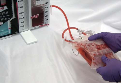
Bottom line: Although shed blood from the chest looks like whole blood, it’s missing key coagulation factors and will not clot. Reinfusing it will boost oxygen carrying capacity, but it won’t help with clotting. You may use it as part of your massive transfusion protocol, but don’t forget to give plasma and platelets according to protocol. This also explains why you don’t need to add an anticoagulant to the autotransfusion unit prior to collecting or giving the shed blood!

Related post: Chest tubes and autotransfusion

Reference: Autotransfusion of hemothorax blood in trauma patients: is it the same as fresh whole blood? Am J Surg 202(6):817-822, 2011.

Print Friendly, PDF & Email

How To See The Unseeable: The Answer

Yesterday I posed a scenario where the surgeon needed to see an area of an open abdomen (trauma laparotomy) that could not easily be visualized. Specifically, there was a question as to whether the diaphragm had been violated just anterior to the liver, just under the costal margin.

Short of putting your head in the wound, how can you visualize this area? Or some other hard to reach spot? Well, you could have an assistant insert a retractor and pull like crazy. However, the rib cage might not bend very well, and in elderly patients it may break. Not a good idea.

Some readers suggested breaking out the laparoscopy equipment and using the camera and optics to visualize. This is a reasonable idea, but expensive. Shouldn’t there be some good (and cheap) way to do this?

Of course, and there is. Think low tech. Very low tech. You just need to see around a corner, right. So get a mirror!

Every OR has some sterile dental mirrors lying around. Get one and have your assistant gently hold the liver down while you indirectly examine the diaphragm. Since you’re probably not a dentist, it may take a minute or two to get used to manipulating the mirror to see just what you want. But if you can manage laparoscopic surgery, you’ll get the hang of it quickly.

And if you need more light up in those nooks and crannies? Shine the OR light directly into the abdomen, then place a nice shiny malleable retractor into the area to reflect light into the area in questions. Voila!

Bottom line: A lot of the things that trauma professionals need to do in the heat of the moment will not be found in doctor, nurse, or paramedic books. Be creative. Look at the stuff around you and available to you. Figure out a way to make it work, and make $#!+ up if necessary.

Print Friendly, PDF & Email

The  May Trauma MedEd Newsletter Is Available!

This month’s newsletter addresses the electronic trauma flow sheet, and more generally, the electronic health record (EHR) in trauma. Here are the topics covered:

  • History Of The Electronic Health Record (EHR)
  • EHRs By The Numbers
  • The EHR And Productivity In The ED
  • Trauma Patient Stay In The ED After EHR
  • The EHR Trauma Flow Sheet
  • What’s The Real Bottom Line?

Subscribers received this issue last week. Subscribe now and be sure to get the next issue early.  So sign up for early delivery now by clicking here!

Click here to download the current issue

Pick up back issues here!

Print Friendly, PDF & Email

Trauma Surgery Tip: How To See The Unseeable

Let me present a scenario and first see how you might solve this problem.

A young man presents with a gunshot to the abdomen in the right mid-back. He is hemodynamically stable, and you get a chest xray. It shows a small caliber slug in the right upper quadrant, but no hemo- or pneumothorax. He has peritoneal signs, so you whisk him off to the OR for a laparotomy.

As you prep the patient for the case, you can feel a small mass just above the right costal margin. You incise the area and produce a 22 caliber bullet. Of course, you follow the chain of evidence rules and pass it off for the police. As you explore the abdomen, it appears that there are no gross injuries. You are concerned, however, that there may be an injury to the diaphragm in proximity to the bullet.

So here’s the question: how can you visualize the diaphragm in this area? The bullet was located below the right nipple. But the diaphragm in this area is covered by the liver, and is parallel to the floor. You can’t seem to feel a hole with your fat finger. But short of putting your whole head in the wound, you just can’t get a good angle to see the area in question.

How would you do it? Please tweet or leave comments with your suggestions. I’ll provide the answer(s) Monday!

Print Friendly, PDF & Email