Category Archives: General

What The Heck? The Answer

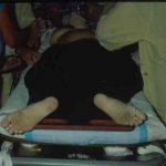
The photo on Friday shows a woman who had been run over by her own car. The vehicle had rolled over her pelvis and stopped, requiring extrication. The most likely injury is an open book pelvic fracture with significant diastasis and/or bilateral unstable sacral fractures.

If you see this clinical presentation there are several things you need to do immediately:

  1. Call for blood. Losses will be large, so you may even want to consider activating your massive transfusion protocol.
  2. Call an orthopedic surgeon. External stabilization will be needed to help decrease blood loss.
  3. Consider early intubation for control of pain. You will be doing a lot, and a patient in agony will slow you down. Your patient is already hypovolemic, so plan your drug choices accordingly.
  4. Search for evidence of an open fracture. Do a good rectal and vaginal exam looking for blood.

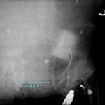
The pelvic xray is poor quality, but shows the major problem, a 10cm pubic diastasis from the open book pelvis fracture. Wrapping the pelvis may be of some help, but consult your orthopedic surgeon first. This pelvis is probably not connected to the spine anymore, so wrapping may have variable results.

Delayed Splenic Rupture: Part 2

Yesterday I wrote about the history of “delayed splenic rupture.” Today I’ll discuss how to deal with it.

If possible, try to avoid ever having to mess around with this clinical problem. If you order an abdominal CT after blunt trauma and see a splenic contrast blush of either type (pseudoaneurysm or extravasation, see left photo), then deal with it before the patient even knows he has a problem. A trip to interventional radiology will usually solve the problem. And if embolized, these patients almost never come back with a bleeding problem.

As I’ve said many times before, if the patient is hemodynamically compromised, then an OR visit is required. The usual solution is splenectomy. Some recommend repairing the spleen, but this is technically more difficult than it sounds, and it is difficult for the surgeon to sleep soundly after performing one of these.

Lets say you inherited one of these from someone else, or ignored the warning signs on the initial CT. The usual time frame for presentation to the ED with acute bleeding is 7 to 10 days after the initial injury. If they are not stable, physical exam or FAST will quickly direct you to the OR, once again for splenectomy. Some patients will stabilize with fluids and can safely be sent to CT scan.

Once the CT confirms what the problem is, a trip to interventional radiology is in order if the patient remains stable. Here is the key: the radiologist must embolize something! If they find a bleeding vessel, then they can selectively embolize it. If they don’t, then the main splenic artery should be embolized. This will decrease the arterial pressure head, but won’t eliminate it. It will decrease the likelihood of additional bleeding as much as possible.

At this point, the patient should be admitted to the trauma service and monitored using your solid organ injury protocol. If they have any hemodynamic issues, it’s time to remove the spleen. Remember, this is the third time they’ve had a problem, and like in baseball, their spleen is out! Attempted splenorrhaphy at this point is pointless and may lead to yet another operation.

Related posts:

Delayed Splenic Rupture: Part 1

This post was prompted by a paper that somehow got into the Journal of Trauma this month on nonoperative management of delayed splenic rupture after trauma. It’s a bad retrospective review of 15 patients which I’ll say more about tomorrow. There’s very little good literature on this topic, so I wanted to share some personal observations.

Back in the days before CT scan (and unfortunately, I remember them), the diagnosis of abdominal injury was much more difficult. It was primarily qualitative, meaning that we somehow figured out that they either had it or they didn’t. We could not very easily figure out what specific injuries a given patient had. However, management was simple: we went to the operating room, found out and fixed it.

Sometimes, though, we would encounter a patient who had been involved in some type of blunt trauma a week or two earlier who presented to the ED with left-sided abdominal pain, shock and anemia. The diagnosis was “delayed splenic rupture” and they were taken to OR for a splenectomy.

When CT scan came along, we found out that these were actually “delayed recognition of splenic injury.” We still took them to the OR for splenectomy, but with experience this slowly gave way to splenic repair, and then to nonoperative management. 

There is still one subset of these injuries that is problematic: spleen injury with a contrast blush. It turns out that there are really two types of blush: contrast seen within a pseudoaneurysm within the splenic pulp, and extravasation. And furthermore, the pseudoaneurysm is the culprit in most “delayed splenic ruptures.”

Tomorrow, I’ll write about how to recognize this potential problem, what to do about it acutely, and what to do if it was missed and the patient presents to your ED ten days later in shock.

Related post:

Reference: Nonsurgical management of delayed splenic rupture after blunt trauma. J Trauma 72(4):1019-1023, 2012.