Category Archives: General

Pop Quiz! Final Answer

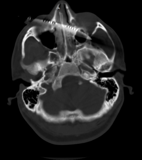
Our patient with the steak knife to the head has been evaluated by CT. The scan shows that the blade enters the right orbit, passing through the medial orbital wall into the ethmoid sinus, turbinates and nasal septum. It then passes into the left orbit along the posterior floor and exits the apex. The optic nerves are not involved, but there may be involvement of the rectus or oblique muscles to the globe. There does not appear to be any involvement of the maxillary sinus.

See why a good exam is important? Gross visual acuity and extra-ocular muscle testing is very important here. Miraculously, all are intact. So now what?

Just yank it out? Absolutely not! Although there is no gross bleeding from the nose or mouth, and none is seen on CT, that doesn’t mean there won’t be! The patient needs to go to the OR, and it may be helpful to have a facial surgeon present just in case. Scopes for evaluating the sinuses and packing materials should be readily available.

Under sedation, the knife can be smoothly withdrawn. An awake patient can tell you how it feels, and whether he is experiencing any bleeding or ocualr changes. If in doubt, the sinuses can be scoped and the globes re-examined.

Note: If troublesome bleeding does occur, this is not an area that is amenable to surgical exploration. The only realistic options available are packing and angioembolization.

Pop Quiz: The Case, Part 3

So our patient has presented to your ED, on foot, with a steak knife sticking out of his head! You’ve activated your trauma team, so now what do you do?

As always, start with a thorough physical exam. A good exam of the head is imperative, as is a scrupulous neurologic exam. In this case, the blade enters just below the right eye, traveling front to back and staying just about level.

Make sure there are no other injuries. Remember the Dang Factor! Don’t focus on the knife and miss other important injuries. And by all means, don’t take it out in the ED!

Since this patient is stable and neurologically intact, the surgeons will want a better idea of the structures involved under the skin. CT is the best tool for this, although there will be scatter from the metal. Here is a representative image:

So now, think about how you will get this out. Tweet and comment your answers.

Related post:

Pop Quiz! The Case, Part 2

Yesterday I presented the case of a young man who shows up at the triage desk in your ED with “something wrong with his head.” I showed an AP skull film, which shows some kind of metallic foreign object. What is it? Where is it? What to do?

First, look at the image carefully. The object is metallic density and appears very thin. But remember, any diagnostic image you view is a 2D representation of a 3D space. You have no idea of the orientation of the object, or exactly where (front to back) it is located. He could be lying on top of it, or it could be stuck in his brain.

At the far left side of the image, the thin metal appears to get even thinner. Dead giveaway! Look at the diagram below.

The knife tang is the thin part of a knife that the handle is fastened to. @andrewjtagg tweeted that he wouldn’t mind seeing a lateral, so here it is.

Yes, it’s a knife. A steak knife to be exact. Somewhere in the middle of the face.

First off, you didn’t need to see these to start doing the right things. Since this is a penetrating injury to the “head, neck or torso” it should trigger any trauma center’s highest level of activation. He is whisked off to the trauma bay and quickly evaluated. He’s obviously awake and alert (he walked in), so what do you need to treat him, and how would you manage it?

Tweet or leave comments. More discussion (and pictures) on Monday.

Pop Quiz! The Case

A young man presents to your emergency department walk-in area. Something is wrong with his head. Here is an AP skull film (when is the last time you got one of those?)

I’ll walk you through my thought processes over the next several days. First, what’s going on? And what should you do now? And next, and so on.

Please tweet and leave comments! My explanation of the initial steps tomorrow.

How To: Secure An Endotracheal Tube To… Nothing!

Several decades ago I took care of a patient who posed an interesting challenge. He had been involved in an industrial explosion and had sustained severe trauma to his face. Although he was able to speak and breathe, he had a moderate amount of bleeding and was having some trouble keeping his airway clear.

Everyone frets about getting an airway in patients who have severe facial trauma. However, I find it’s usually easier because the bones and soft tissue move out of your way. That is, as long as you can keep ahead of the bleeding to see your landmarks.

In this case, the intubation was easy. The epiglottis was visible while standing above the patient’s head, so a laryngoscope was practically unnecessary! But now, how do we secure the tube so it won’t fall out? Sure, there are tube-tamer type securing devices available, but what if they are not available to you? Or this happened in the field? Or it was in the 1980’s and it hadn’t been invented, like this case?

The answer is, create your own “skin” to secure the tube to. Take a Kerlix-type stretchable gauze roll and wrap it tightly around their head. Remember, they are sedated already and they can breathe through the tube. This also serves to further slow any bleeding from soft tissue. Once you have “mummified” the head with the gauze roll, tape the tube in place like you normally would, using the surface of the gauze as the “skin.”

Be generous with the tape, because the tube is your patient’s life-line. Now it’s time for the surgeons to surgically stabilize this airway, usually by converting to a tracheostomy. 

Related posts: