Category Archives: Complications

Treatment Of BCVI

In my last post, I reviewed the grading system for blunt carotid and vertebral artery injury (BCVI). Today, we’ll wrap up and discuss treatment.

There are basically three modalities at our disposal for managing BCVIantithrombotic medication (heparin and/or antiplatelet agents), surgery, and endovascular procedures. The choice of therapy is usually based on surgical accessibility and patient safety for anticoagulation. We do know that a number of studies have shown a decrease in stroke events in patients who are heparinized. Unfortunately, this is not always possible due to associated injuries. Antiplatelet agents are usually tolerated after acute trauma, especially low-dose aspirin. Several studies have shown little difference in outcomes in patients receiving heparin vs aspirin/clopidogrel for BCVI.

So what to do? Here are some broad guidelines:

  • Grade I (intimal flap). Heparin or antiplatelet agents should be given. If heparin can be safely administered, it may be preferable in patients who will need other surgical procedures since it can be rapidly reversed just by stopping the infusion. These lesions generally heal completely on their own, so a followup CT angiogram should be scheduled in 1-2 weeks. Medication can be stopped when the lesion heals.
  • Grade II (flap/dissection/hematoma). These injuries are more likely to progress, so heparin is preferred if it can be safely given. Stenting should be considered, especially if the lesion progresses. Long-term anti-platelet medication may be required.
  • Grade III (pseudoaneurysm). Initial heparin therapy is preferred unless contraindicated. Stable pseudoaneurysms should be followed with CTA every 6 months. If the lesion enlarges, then surgical repair should be carried out in accessible injuries or stenting in inaccessible ones.
  • Grade IV (occlusion). Heparin therapy should be initiated unless contraindicated. Patients who do not suffer a catastrophic stroke may do well with followup antithrombotic therapy. Endovascular treatment does not appear to be helpful.
  • Grade V (transection with extravasation). This lesion is frequently fatal, and the bleeding must be addressed using the best available technique. For lesions that are surgically accessible, the patient should undergo the appropriate vascular procedure. Inaccessible injuries should undergo angiographic treatment and may require embolization to control bleeding without regard for the possibility of stroke.

References:

  1. Scott WW, Sharp S, Figueroa SA, et al. Clinical and radiographic outcomes following traumatic Grade 1 and 2 carotid artery injuries: a 10-year ret-rospective analysis from a Level I trauma center. J Neurosurg 122:1196, 2015.
  2. Scott WW, Sharp S, Figueroa SA, et al. Clinical and radiographic outcomes following traumatic Grade 3 and 4 carotid artery injuries: a 10-year ret-rospective analysis from a Level 1 trauma center. J Neurosurg 122:610, 2015.
  3. Scott WW, Sharp S, Figueroa SA, et al. Clinical and radiological outcomes following traumatic Grade 1 and 2 vertebral artery injuries: a 10-year retrospective analysis from a Level 1 trauma cen-ter. J Neurosurg 121:450, 2015.
  4. Scott WW, Sharp S, Figueroa SA, et al. Clinical and radiological outcomes following traumatic Grade 3 and 4 vertebral artery injuries: a 10-year retrospective analysis from a Level I trauma center. The Parkland Carotid and Vertebral Artery Injury Survey. J Neurosurg 122:1202, 2015.
Print Friendly, PDF & Email

How To Grade BCVI

In my last post, I reviewed the three screening systems for blunt carotid and vertebral artery injury (BCVI). Today, we’ll look at grading them.

Only 10 years ago, it was a major production to identify BCVI. CT angiography was still in its early days, and scanner resolution and radiologist experience were major issues that impacted accuracy.

We’ve come a long way in a relatively short period of time, and current day scanners now have more than adequate resolution. It’s also more common to have a radiologist with special skills reading these studies, the neuroradiologist. For these reasons, CT angiography has become the standard for diagnosis. It is also the most cost-effective. Only in very rare cases do we need to obtain a conventional contrast angiogram.

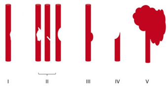
Once the study has been obtained, it’s time to identify and classify the injury. The Denver group is also responsible for bringing us the grading system for BCVI. See the diagram below.

Here are the details:

Grade I: A mild intimal irregularity is seen. Note the abnormally narrowed area, representing a small intimal injury, possibly with a small amount of clot.

Grade II: This grade has several presentations. There may be an intraluminal thrombosis/hematoma with (left) or without (right) an intimal flap, or a flap alone (center).

Grade III: There is a full-thickness injury to the vessel with a contained extraluminal extravasation (pseudoaneurysm)

Grade IV: The vessel is completely occluded by flap or thrombus

Grade V: The artery is transected and freely extravasating

Here’s a nice diagram:

Remember, we always grade things for a reason! Ultimately, the injury grade will translate into the selection of treatment. We’ll cover that in my next post.

Reference: Blunt carotid arterial injuries: implications of a new grading scale. J Trauma. 1999;47(5): 845-53.

Print Friendly, PDF & Email

Screening For BCVI

In my last post, I described how common we think blunt carotid and vertebral injury (BCVI) really is. Today, I’ll review how we screen for this condition.

Currently, there are three systems in use: Denver, Expanded Denver, and Modified Memphis. Let’s look at each in detail.

Denver BCVI Screening

There is an original Denver screening system, and a more recent modification. The original system was divided into mechanism, physical signs, and radiographic findings. It was rather rudimentary and evolved into the following which uses both signs and symptoms, and high-risk factors.

Signs and symptoms

  • potential arterial hemorrhage from the neck, nose, or mouth
  • cervical bruit in patients <50 years of age
  • expanding cervical hematoma
  • focal neurologic deficit (transient ischemic attack, hemiparesis, vertebrobasilar symptoms, Horner syndrome) incongruous with head CT findings
  • stroke on CT

Risk factors

  • Le Fort II or III mid-face fractures
  • Cervical spine fractures (including subluxations), especially fractures involving transverse foramen or C1-C3 Vertebrae
  • Basilar skull fracture and involvement of carotid canal
  • Diffuse axonal injury with GCS <8
  • Near hanging with anoxic brain injury
  • Seat belt sign (or other soft tissue neck injury) especially if significant associated swelling or altered level of consciousness

The Denver group reviewed their criteria in 2012 and found that 20% of the patients who had identified BCVI did not meet any of their criteria. And obviously, this number cannot include those who were never symptomatic and therefore never discovered.

Based on their analysis, they added several additional risk factors to the original system:

  • Mandible fracture
  • Complex skull fracture/basilar skull fracture/occipital condyle fracture
  • TBI with thoracic injuries
  • Scalp degloving
  • Thoracic vascular injuries
  • Blunt cardiac rupture

The downside of these modifications is that they are a little more complicated to identify. The original criteria were fairly straightforward yes/no items. But “TBI with thoracic injuries?” Both the TBI part and the thoracic injury part are very vague. This modification casts a wider net for BCVI, but the holes in the net are much larger.

Memphis BCVI Screening

Let’s move on to the modified Memphis system for identifying BCVI. It consists of seven findings that overlap significantly with the Denver criteria. The underlined phrases indicated the modifications that were applied to the original criteria.

  • base of skull fracture with involvement of the carotid canal
  • base of skull fracture with involvement of petrous temporal bone
  • cervical spine fracture (including subluxation, transverse foramen involvement, and upper cervical spine fracture)
  • neurological exam findings not explained by neuroimaging
  • Horner syndrome
  • Le Fort II or III fracture pattern
  • neck soft tissue injury (e.g. seatbelt sign, hanging, hematoma)

Interestingly, these modifications were first described in an abstract which was never published as a paper. Yet somehow, they stuck with us.

So there are now two or three possible systems to choose from when deciding to screen your blunt trauma patient. Which one is best?

Let’s go back to the AAST abstract presented by the Birmingham group this year that I mentioned previously. Not only did they determine a more accurate incidence, but they also tested the three major screening systems to see how each fared. See Table 1.

Look at these numbers closely. When any of these systems were applied and the screen was negative, the actual percentage of patients who still actually had the injury ranged from about 25% to 50%! Basically, it was a coin toss with the exception of the Expanded Denver criteria performing a little better.

If you are a patient and you actually have the injury, how often does any screening system pick it up? Oh, about one in five times. Again, this is not what we want to see.

So what to do? The Expanded Denver screen has a lower false negative rate, but the total number of positive screens, and hence the number of studies performed, doubles when it is used.

Here’s how I think about it. BCVI is more common than we thought in major blunt trauma. If not identified, a catastrophic stroke may occur. Current screening systems successfully flag only 50% of patients for imaging. So in my opinion, we need to consider imaging every patient who is already slated to receive a head and cervical spine CT after major blunt trauma! At least until we have a more selective (and reliable) set of screening criteria.

References:

  • (Denver) Optimizing screening for blunt cerebrovascular injuries. Am J Surg. 1999;178:517–522.
  • (Expanded Denver) Blunt cerebrovascular injuries: Redefining screening criteria in the era of noninvasive diagnosis. J Trauma 2012;72(2):330-337.
  • (Memphis) Prospective screening for blunt cerebrovascular injuries: analysis of diagnostic modalities and outcomes. Ann Surg. 2002, 236 (3): 386-393.
  • (Modified Memphis) Diagnosis of carotid and vertebral artery injury in major trauma with head injury. Crit care. 2010;14(supp1):S100.
Print Friendly, PDF & Email

Why Do Trauma Patients Get Readmitted?

Readmission of any patient to the hospital is considered a quality indicator. Was the patient discharged too soon for some reason? Were there any missed or undertreated injuries? Information from the Medicare system in the US (remember, this represents an older age group than the usual trauma patient) indicates that 18% of patients are readmitted and 13% of these are potentially preventable.

A non-academic Level II trauma center in Indiana retrospectively reviewed their admissions and readmissions over a 3 year period and excluded patients who were readmitted on a planned basis (surgery), with a new injury, and those who died. This left about 5,000 patients for review. Of those, 98 were identified as unexpected readmissions. 

There were 6 major causes for readmission:

  • Wound (23) – cellulitis, abscess, thrombophlebitis. Two thirds required surgery, and 4 required amputation. All of these amputations were lower extremity procedures in obese or morbidly obese patients.
  • Abdominal (16) – ileus, missed injury, abscess. Five required a non-invasive procedure (mainly endoscopy). Only 2 required OR, and both were splenectomy for spleen infarction after angioembolization.
  • Pulmonary (7) – pneumonia, empyema, pneumothorax, effusion. Two patients required an invasive procedure (decortication, tube placement).
  • Thromboembolic (4) – DVT and PE.  Two patients were admitted with DVT, 2 with PE, and 1 needed surgery for a bleed due to anticoagulation.
  • CNS (21) –  mental status or peripheral neuro exam change. Eight had subdural hematomas that required drainage; 3 had spine fractures that failed nonoperative management.
  • Hematoma (5) – enlargement of a pre-existing hematoma. Two required surgical drainage.

About 14% of readmissions were considered to be non-preventable by a single senior surgeon. Wound complications had the highest preventability and CNS changes the lowest. Half occurred prior to the first followup visit, which was typically scheduled 2-3 weeks after discharge. This prompted the authors to change their routine followup to 7 days.

Bottom line: This retrospective study suffers from the usual weaknesses. However, it is an interesting glimpse into a practice with fewer than the usual number patients lost to followup. The readmission rate was 2%, which is pretty good. One in 7 were considered “preventable.” Wounds and pulmonary problems were the biggest contributors. I recommend that wound and pulmonary status be thoroughly assessed prior to discharge to bring this number down further. Personally, I would not change the routine followup date to 1 week, because most patients have far more complaints that are of little clinical importance than compared to 2 weeks after discharge.

Reference: Readmission of trauma patients in a nonacademic Level II trauma center. J Trauma 72(2):531-536, 2012.

Print Friendly, PDF & Email

Fatigue V: Your Patients

I’ve spent the past several posts detailing how interrupted sleep interferes with the health and effectiveness of trauma professionals. But what about our patients? Being in the hospital is nothing like trying to sleep at home. The beds are terrible. There is noise in the hall. Their roommate is confused and calls out at all hours. Nurses keep coming in to check vital signs. The pulse oximeter beeps every 10 minutes.

How bad is it, really? There is no trauma patient-specific literature yet on the topic. But there is a recently published paper detailing the experience of general surgery patients admitted after elective procedures that is very revealing. The group at Dartmouth-Hitchcock Medical Center recruited adults who stayed at least one night at the hospital.

Prior to surgery, each patient completed a questionnaire that measured their baseline home sleep quality. Postop, they completed another questionnaire designed to measure their in-hospital sleep quality.  Each patient was fitted with a Fitbit Inspire HR tracker, which they wore during their entire hospitalization.  After discharge home, they completed a final outpatient sleep questionnaire.

Here are the factoids:

  • A total of 74 patients were recruited and 54 completed all phases of the study; 59 finished all of the pre-discharge phases
  • The average inpatient sleep score was 49/100, where scores less than 50 are considered substandard
  • The major culprit for in-hospital sleep disturbance was nighttime awakenings
  • Patients who had better home sleep quality tended to have a higher in-hospital score (65)
  • Sleep quality was so poor that only 40% of Fitbit devices were able to record sleep time on the first postop night, and that average time asleep was 4.7 hours
  • As expected, patients with a roommate did not sleep as well as those in private rooms
  • Average sleep time increased over subsequent nights to about 6 hours, which is still short of the recommended 7 hour minimum
  • About 88% of patients were poor sleepers preop (!), and this did not change after they returned home (85%)

Bottom line: Sleep quality in the hospital is terrible! I can vouch for this from the standpoint of being a surgeon on call, and also from one experience as a patient. There is very good data on the adverse effects of sleep loss. Fasting glucose and systolic blood pressure rise significantly after a single night of poor sleep. When these occur in a hospital, this sets the clinicians into a frenzy of prescribing sliding scale insulin or antihypertensives and other meds that are probably not needed.

There are numerous other more subtle effects as well. The best way to avoid them is to promote good sleep. But how can you do this in the hospital? Here are some tips:

  • Be aware that the way you order medications (tid vs q8hrs) has a big impact on when the evening/night doses are given. Talk to your pharmacist so your patient only gets meds when they would normally be awake and not in the middle of the night.
  • Remove unnecessary monitors that might alarm during sleep. Pulse oximeters are probably the biggest culprit. Does it need to be continuous, or can (very) occasional spot checks be done?
  • Does your patient really need vital signs taken during the middle of the night? After the first few shifts, most ward patients do not.
  • Watch out for the phlebotomists! They love to circulate early so lab results are available at the crack of dawn. Can’t it wait?
  • Some practice guidelines call for repeat scans or other studies after a certain number of hours. If one is due at 3am, can’t it wait until morning? Really?
  • On a daily basis, review all actual or potential nighttime interruptions with the patient and their nurse. Discontinue or reschedule anything that really can’t wait until morning.

Failing to provide for good sleep quality sets your patient up for complications, abnormal vitals and blood tests, and altered mental status. Do everything you can to optimize their sleep!

Reference: Deep sleep and beeps: sleep quality improvement project in general surgery patients. JACS 232(6):882-888, 2021.

Print Friendly, PDF & Email