Category Archives: Abdomen

Nonoperative Management Of Abdominal Stabs: How Long Should We Watch?

Gunshots to the abdomen are a no-brainer that nearly always require operative exploration, but stab wounds are more challenging. They are low velocity, and injury only occurs in the pathway of the knife. It is more likely that inconsequential (or no) injuries occurred. Since exploratory laparotomy (or even laparoscopy) is not a benign procedure, trauma professionals frequently opt for selective nonoperative management (observation) in these cases.

What does observation mean? The patients are kept in the hospital for a set period of time, receive serial abdominal examinations, and get a few repeat lab tests. If the exam changes, the patient is taken to the OR to find out why. If it doesn’t, they are fed and sent home.

But what is the appropriate period of time to observe? One major concern is for the possible hollow viscus injury. Stomach and colon contain fluids that cause prompt peritonitis. But small intestinal content is rather innocuous, with neutral pH, normal concentration, and few bacteria. Peritonitis may not occur for days. Yet most centers send these patients home within 12 to 24 hours of injury.

What is the right answer? The trauma group at LAC+USC in Los Angeles performed a prospective, observational study to try to answer the question. They enrolled all patients with abdominal stab wounds presenting to their center over a three year period. They were generous (and correct) with their definition of the abdomen, including the thoracoabdominal portion up to the 5th intercostal spaces, and the pelvis.

Patients were excluded if they were hemodynamically unstable, or had an evisceration or obvious peritonitis. Most patients received a CT scan, and patients with suspected hollow viscus injury were excluded from the study and taken to the OR. All others were observed in a dedicated unit and were monitored for change in exam, need for blood transfusion, or other event which was recorded for the study.

Here are the factoids:

  • A total of 256 patients met study criteria: 77% had a single stab and 76% had a negative FAST exam
  • There were 46 patients who underwent immediate laparotomy for evisceration (59%), hemodynamic instability (33%), or peritonitis (24%)
  • 81% of patients underwent CT scan, and 13% were taken to OR based on the findings
  • This left 210 patients for nonoperative management
  • Of these, 71 had positive scans and all were due to solid organ injury. One patient failed at 32 hours due to increasing lactate and WBC, but the operation was nontherapeutic.
  • Another 14 patients had equivocal CT findings and two failed at 10 and 20 hours due to small bowel injury discovered by increasing lactate, WBC, change in exam, and air on a repeat CT
  • All 123 patients with negative scans passed nonoperative management
  • Median hospital length of stay was 3 with a range of 2-6 days

Since all of their patients who failed observation did so within 24 hours, the authors recommended a 24 hour observation period for all patients with stabs to the abdomen who did not meet their exclusion criteria.

Bottom line: Trauma professionals have needed a study like this for decades. Until now, we’ve been flying by the seat of our pants, with each surgeon making up his or her own magic number. This is a well done first attempt at defining what that number should be. 

Yes, there are some limitations to the study. The most important one that we don’t know the answer to is how many patients were successfully discharged that presented to another hospital with complications or failure. But this study provides a very reasonable estimate that helps us balance the cost (and patient inconvenience) of time in the hospital vs the dangers of a delayed diagnosis.

The only thing that remains is to design the practice guideline that incorporates the observation period, how often serial exams and labs should be obtained, and when the CT should be repeated.

Reference: Prospective evaluation of the selective nonoperative management of abdominal stab wounds: When is it safe to discharge? Journal of Trauma and Acute Care Surgery: November 2022 – Volume 93 – Issue 5 – p 639-643.

Trauma And The Gut Microbiome

This is a follow-on post from one published last week (gut microbiome changes in rats due to trauma). Click here to read it.

One of the newest frontiers in health-related research recognizes the importance of the human microbiome. This term describes the collection of all genomes from the microorganisms found in a particular environment, such as in, on, or around a human. The term microbiota refers to the specific bacteria, viruses, and fungi that colonize the areas within this environment.

Within the last decade or so, we have just begun to appreciate the importance of the microorganisms that live within us. From a purely numerical standpoint, there are 10 times as many of them as there are our own human cells. However, since they are so small in comparison, we can’t really appreciate the huge number of “other” cells in and on us.

These tiny cohabitants provide many, many functions that are important to our health and well-being. They protect us from pathogenic organisms, help digest our food, fine-tune our immune system, and synthesize proteins, amino acids, and vitamins that are essential to our health. And much more!

The usual microbiota can be disrupted by disease, poor diet, stress, and even a single dose of antibiotics. With each new research paper, we recognize new functions for and disruptors of our microbiota.

The surgery groups at two San Antonio hospitals, UT Health and the US Army Institute of Surgical Research, recognize the importance of the gut microbiota, building upon prior work demonstrating changes within it in the presence of trauma and burn injury.

The authors performed a prospective, observational cohort study of severely injured patients over a two-year period. They attempted to characterize differences in the microbiota between trauma patients and to identify changes in these communities over time.

A rectal swab was obtained from each patient shortly after admission and the microbial DNA present was identified. This was repeated regularly throughout the hospital stay.

Here are the factoids:

  • 72 patients and 13 healthy controls were enrolled
  • Patients were severely injured with a mean ISS of 21; an average of 6 units of blood products were given in the first 24 hours
  • Only one fourth of the injured patients had a microbiome similar to the healthy controls
  • These 26% received significantly more blood products than the dissimilar patients (14 units vs 3)
  • There were significant decreases in the numbers and ratios of normal gut bacteria and increases in the numbers of pathogenic bacteria. These changes increased with time in hospital

Bottom line: Yes, this is new and bizarre territory. It appears that shock, hypoxia, medications (and not just antibiotics), surgical intervention, and poor nutrition can adversely affect the microflora in our gut. Conversely, early transfusion seems to ameliorate this effect to some degree.

At this point in time, there is nothing you can do with this knowledge. Just be aware that everything you routinely do to your patients can change their microbiota, and this may in turn have unexpected effects on their health and recovery. I anticipate seeing many more papers like this one in the near future.

Reference: A prospective study in severely injured patients reveals an altered gut microbiome is associated with transfusion volume. J Trauma 86(4):573-582, 2019.

Surveillance For Splenic Pseudoaneurysm After Injury

When it comes to repeat CT scanning after splenic injury, there are believers and there are non-believers. In my experience, the majority of centers in the US are non-believers. However, there is a new paper in press that attempts to convince us that more should become believers.

I think the biggest lesson to be learned from this paper is that WE SHOULD READ THE ENTIRE PAPER before drawing conclusions. I have said this in the past and I will say it again. In this case, not only did I read the entire paper, but I had to dig deep into the references it cited as well.

Nonoperative management of splenic injuries has a very high success rate if done properly. Some papers claim this can be up to 93%, which parallels my experience. This success rate involves excluding unstable patients (they need to be in the operating room) and planned use of angioembolization in select patients. Over the years we have found that we need to do less and less in the management of solid organ injury patients:

  • No bedrest
  • No starvation (NPO status)
  • No serial blood draws
  • No repeat CT scan
  • Few limitations on activity after discharge

For an example of a practice guideline that demonstrates that less is more, use the download link at the end of this post.

But back to the question about repeat CT scanning before discharge. Why do we need to do this? The usual reason is that “we want to find delayed pseudoaneurysms.” And why is that important? “It might bleed!”

Really? Let’s look into that through the lens of this new paper by the group at the University of Cincinnati. They performed a retrospective study of their experience with patients who had sustained blunt splenic injury during a recent three-year period. They were interested in how many underwent splenectomy or splenorrhaphy, who had repeat CT imaging, who went to interventional radiology (IR) and when, and which ones were found to have pseudoaneurysms and what was done about it.

Here are the factoids:

  • There were 539 patients who met inclusion criteria, with an average ISS of 24
  • Of these, 46 died during their hospital stay (none from their splenic injury)
  • Focusing on the 248 patients with higher grade injuries (III-V), 125 (50%) underwent emergent or delayed splenectomy. Early vs late operation was not broken out, but this is a startlingly high number!
  • Of the higher grade injured patients who kept their spleens, 97% underwent repeat CT around day 5
  • Delayed pseudoaneurysms were detected in the following patients:
    • Grade III: 10 of 88 patients (11%). Then 8 of those 10 went to IR, and 5  of 10 had splenectomy!
    • Grade IV: 7 of 24 (29%).  Then 8 of the 7 (error in the paper?) went to IR and 3 of 7 had splenectomy!
    • Grade V: 2 of 5 (40%). Both of these patients went to IR and somehow kept their spleens.

The authors conclude that routine followup CT imaging identifies splenic pseudoaneurysms allowing for interventions to minimize delayed complications.

Bottom line: Whoa! There’s a lot going on here. My first observation is that this center does a lot of splenectomies! Of the 539 patients (all comers) who were included in the study, 129 (24%)  lost their spleens. If grade I-II injuries are excluded that percent rises to 50%!

Only eight splenectomies were performed after the repeat CT. This would imply that there were either a lot of unstable patients with splenic injury, the institutional indications for this procedure arbitrarily include grade, or there is a lot of variability in the decision to perform it.

I think there are really two questions to answer here. 

  1. Does delayed splenic pseudoaneurysm occur? The answer is yes. There are a few studies (performed by believers) that demonstrate new pseudoaneurysms after repeat CT. I’m convinced.
  2. Do we care? The real question is, do these pseudoaneurysms cause harm? The fear is that they might explode at some point after patient discharge and cause a major problem.

Papers written by the believers cite a number of old studies and give numbers between 2% and 27% for incidence of delayed hemorrhage. Well, I tracked down all of these papers, including the ones they cited. And it doesn’t add up.

  • One paper from a believer institution found no delayed bleeds.
  • Several papers were for pediatric patients, whose spleens don’t behave like adult ones. They found one case after discharge in one out of 276 patients across three studies.
  • Of 76 adolescents, none encountered delayed bleeds

Many of the papers cited regarding bleeding complications are very old. CT scanners had less resolution, and in many papers, IR was not even a consideration. 

So here’s my take. Yes, delayed pseudoaneurysms occur. In children we don’t care. They almost never cause a problem. But in adults, they can and do cause issues and should be embolized shortly after the initial scan. 

Once embolized, the ones seen on that initial scan are effectively neutralized and do not need a repeat scan. The small ones that might pop up later may very well be part of the healing process. And they may not even occur if angioembolization is done early. It seems unlikely that anything further is needed.

But remember, clinical judgement trumps all. If your patient starts complaining of new abdominal symptoms while in the hospital or after discharge, get a prompt CT scan to rule out any developing complications.

Sample solid organ injury protocol: click here

Reference: Delayed splenic pseudoaneurysm identification with surveillance imaging. J Trauma Acute Care Surg. 2022 Mar 22. doi: 10.1097/TA.0000000000003615. Epub ahead of print. PMID: 35319540.


Options For Hemorrhage Control From Pelvic Fracture

We’ve come a long way in our available treatments to slow or stop bleeding from pelvic fractures. Let’s work our way through the list in today’s post, then look critically at two of the newcomers in the next one.

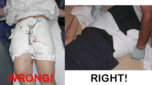
Pelvic binders. Long ago, these were just sheets wrapped around the patient and secured with clamps.

They were rather crude, as you can see. So of course, several enterprising companies began to offer commercial binders that were easier to place and secure.

Of note in the photo above, the wrap on the left is totally wrong. It is too wide and extends too high, so will not provide effective compression. The image on the right shows proper placement low across the greater trochanters. It is also not secured using metal clamps which may interfere with x-ray imaging.

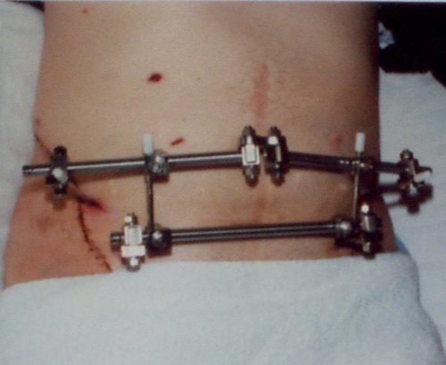
External fixation of the pelvis. This usually involved a call to your friendly orthopedic surgeon. It could be applied in either the trauma bay or the operating room.

This image also shows improper technique. The horizontal bar should be angulated downwards over the pubis so it will not interfere with the trauma surgeon’s approach to laparotomy.

Internal pelvic packing + internal iliac artery ligation. Since surgeons didn’t have many other good tools, they could actually operate! Unfortunately, neither of these worked terribly well. The laparotomy pads could decompress upwards out of the pelvis and the internal iliac arteries have lots of collateral branches that permit ongoing bleeding from pelvic bones.

Angioembolization. Arterial bleeding from the pelvis occurs more often than you think (upwards of 50% of major pelvic injuries). Angiography and embolization can work very well. Unfortunately they are not suitable for unstable patients since IR suites are poor resuscitation areas. Many trauma centers do not have hybrid operating rooms where hemodynamically compromised patients can be taken for combined IR and open procedures if needed. So unstable patients must go to a regular OR first to attempt stabilization.

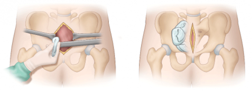
Preperitoneal packing. This is the new OR procedure kid on the block. Instead of placing packs in the pelvis, they are placed next to the broken pelvic bones but just outside the peritoneum. This permits better tamponade, and the intraperitoneal viscera push out against the packs to help decrease bleeding.

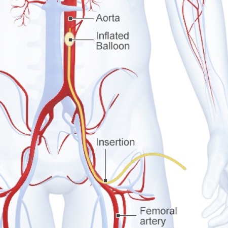
Zone 3 REBOA. And this is the very newest kid on the block. The balloon tipped catheter is inserted to a level above the aortic bifurcation but below the visceral and renal vessels. This is essentially a non-selective, temporary ligation of not just the internal iliac arteries, but everything distal to the aorta. It can be performed in the ED to dramatically slow blood loss, providing more time to get the patient to the OR where more definitive hemorrhage control can be provided (using many of the above techniques).

In my next post, I’ll take a closer look at the effectiveness of preperitoneal packing vs angioembolization.

Do We Really Need To Admit Children With Low-Grade Solid Organ Injury?

Over the years, we have slowly gotten wiser about solid organ injuries (SOI). Way back when, before CT and ultrasound, if there was a suspicion a patient had such an injury you were off to the operating room. We learned (from children, I might add) that these injuries, especially the minor ones, were not such a big deal.

However, we routinely admit adults and children with solid organ injury of any grade. Many centers have streamlined their practice guidelines so that these patients don’t spend very long in the hospital, but most are still admitted. A number of researchers from Level I pediatric centers in the US got together to see if this is really necessary.

They combed through 10 years worth of TQIP data for outcomes and timing of intervention in children with low-grade (grades 1 and 2) solid organ injury age 16 or less. Children with “trivial” extra-abdominal injuries were included to make the conclusions more generalizable.  Penetrating injuries and burns were excluded, as were those with “risk of hemorrhage” or need for abdominal exploration for reasons other than the SOI. The risk of hemorrhage was defined as a pre-existing condition or other injury that made it more likely that a transfusion might be necessary for other causes.

Here are the factoids:

  • A total of 1,019 children with low-grade SOI (liver, kidney, or spleen) were enrolled in the study, and 97% were admitted
  • There was an even distribution across age groups. Many studies over-represent teenagers; this was not the case here.
  • Median LOS was 2 days, and a quarter were admitted to the ICU
  • Only 1.7% required an intervention, usually on the first hospital day (transfusion, angiography, or laparotomy)
  • Pediatric trauma centers did not perform any of the 9 angiographic procedures, and they only performed 1 laparotomy of the 4 reported

The authors concluded that practice guidelines should be developed for adult centers caring for children to decrease the number of possibly unnecessary interventions, and that it may be feasible to manage many children with low-grade SOI outside of the hospital.

Bottom line: This is an intriguing study. The admission length and silly restrictions like bed rest, NPO, and multiple lab draws are finally approaching their end. Although this paper does have the usual limitations of using a large retrospective database, it was nicely done and thoughtfully analyzed. 

It confirms that adverse events in this population are very uncommon, and that adult centers are still too aggressive in treating children like adults. The recommendation regarding practice guidelines is very poignant and this should be a high priority.

Individual centers should determine if they have the infrastructure to identify low-risk children who have reliable families and live in proximity to a hospital with a general surgeon, or better yet, near a trauma center. Hopefully this study will help accelerate the adoption of such guidelines and practices, moving treatment for many children to the outpatient setting.

Reference: Hospital-based intervention is rarely needed for children with low-grade blunt abdominal solid organ injury: An analysis of the Trauma Quality Improvement Program registry. JTrauma 91(4):590-598, 2021.