All posts by The Trauma Pro

Pop Quiz! The Case, Part 2

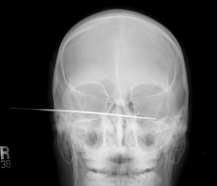
Yesterday I presented the case of a young man who shows up at the triage desk in your ED with “something wrong with his head.” I showed an AP skull film, which shows some kind of metallic foreign object. What is it? Where is it? What to do?

First, look at the image carefully. The object is metallic density and appears very thin. But remember, any diagnostic image you view is a 2D representation of a 3D space. You have no idea of the orientation of the object, or exactly where (front to back) it is located. He could be lying on top of it, or it could be stuck in his brain.

At the far left side of the image, the thin metal appears to get even thinner. Dead giveaway! Look at the diagram below.

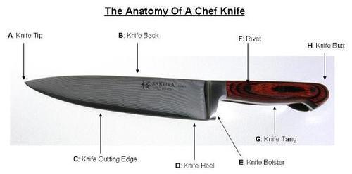
The knife tang is the thin part of a knife that the handle is fastened to. @andrewjtagg tweeted that he wouldn’t mind seeing a lateral, so here it is.

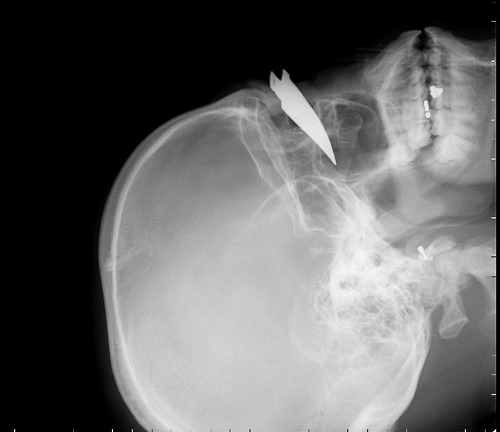
Yes, it’s a knife. A steak knife to be exact. Somewhere in the middle of the face.

First off, you didn’t need to see these to start doing the right things. Since this is a penetrating injury to the “head, neck or torso” it should trigger any trauma center’s highest level of activation. He is whisked off to the trauma bay and quickly evaluated. He’s obviously awake and alert (he walked in), so what do you need to treat him, and how would you manage it?

Tweet or leave comments. More discussion (and pictures) on Monday.

Pop Quiz! The Case

A young man presents to your emergency department walk-in area. Something is wrong with his head. Here is an AP skull film (when is the last time you got one of those?)

I’ll walk you through my thought processes over the next several days. First, what’s going on? And what should you do now? And next, and so on.

Please tweet and leave comments! My explanation of the initial steps tomorrow.

How To: Secure An Endotracheal Tube To… Nothing!

Several decades ago I took care of a patient who posed an interesting challenge. He had been involved in an industrial explosion and had sustained severe trauma to his face. Although he was able to speak and breathe, he had a moderate amount of bleeding and was having some trouble keeping his airway clear.

Everyone frets about getting an airway in patients who have severe facial trauma. However, I find it’s usually easier because the bones and soft tissue move out of your way. That is, as long as you can keep ahead of the bleeding to see your landmarks.

In this case, the intubation was easy. The epiglottis was visible while standing above the patient’s head, so a laryngoscope was practically unnecessary! But now, how do we secure the tube so it won’t fall out? Sure, there are tube-tamer type securing devices available, but what if they are not available to you? Or this happened in the field? Or it was in the 1980’s and it hadn’t been invented, like this case?

The answer is, create your own “skin” to secure the tube to. Take a Kerlix-type stretchable gauze roll and wrap it tightly around their head. Remember, they are sedated already and they can breathe through the tube. This also serves to further slow any bleeding from soft tissue. Once you have “mummified” the head with the gauze roll, tape the tube in place like you normally would, using the surface of the gauze as the “skin.”

Be generous with the tape, because the tube is your patient’s life-line. Now it’s time for the surgeons to surgically stabilize this airway, usually by converting to a tracheostomy. 

Related posts:

The Newest Trauma MedEd Newsletter Is Available!

The April newsletter is now available! Click the image below or the link at the bottom to download. This month’s topic is Protocols (again). You’ll need a QR code reader if you want to download to your mobile device. Or just use the web URLs provided to download to your desktop/notebook.

In this issue you’ll find articles on:

  • Chest tube management
  • Solid organ injury
  • Rapid reversal of warfarin
  • Reversal of other anticoagulants
  • Massive transfusion

Subscribers had the newsletter emailed to them over the weekend. If you want to subscribe (and download back issues), click here.

Download the newsletter here!

Trauma Pearl: Unexpected Respiratory Failure After Blunt Trauma

A 24 year old restrained female is involved in a T-bone type motor vehicle crash. She sustains a moderate to severe traumatic brain injury and is intubated and sedated. On exam, she has a few abrasions over her left flank, and no other physical findings. Head CT shows some subarachnoid blood, and abdominal CT is negative.

She is placed in the ICU and slowly becomes more responsive. However, her FIO2 has to be increased several times due to poor oxygenation. By day 3, she is on 90% O2 and has diffuse infiltrates in her lung fields.

What’s the problem?!

This is a classic presentation of a missed abdominal injury. Restrained patients are at risk for intestinal injuries, even with a t-bone mechanism and little to no seat belt sign. Physical exam may be helpful, but abdominal pain/tenderness may be masked by head injury.

A repeat CT scan was performed, which showed free fluid and a few bubbles of free air. The patient was taken to the OR and a bucket handle injury to the distal ileum was found, with devitalized and leaking intestine. This was resected and primary anastomosis was performed. Within 2 days, the patient was on 40% O2 and was ready for extubation two days later.

Bottom line: Unexplained respiratory failure after blunt trauma, especially if no chest injury has occurred, is nearly always due to a missed abdominal injury. The initial CT is a snapshot that is valid for only a few hours. Re-image with CT or ultrasound, and operate promptly if any significant change in patient condition occurs.


Related posts:

Fictional case, not treated at Regions Hospital.