How To: The Serial Abdominal Exam

How often have you seen this in an admitting history and physical exam note? “Admit for observation; serial abdominal exams.” We say it so often it almost doesn’t mean anything. And during your training, did anyone really teach you how to do it? For most trauma professionals, I believe the answer is no.

Yet the serial abdominal exam is a key part of the management of many clinical issues, for both trauma patients as well as those with acute care surgical problems.

Here are the key points:

  • Establish a baseline. As an examiner, you need to be able to determine if your patient is getting worse. So you need to do an initial exam as a basis for comparisons.
  • Pay attention to analgesics. Make sure you know what was given last, and when. You do not need to withhold pain medications. They will reduce pain, but not eliminate it. You just need enough information to determine if the exam is getting worse with the same amount of medication on board.
  • Perform regular exams. It’s one thing to write down that serial exams will be done, but someone actually has to do them. How often? Consider how quickly your patient’s status could change, given the clinical possibilities you have in mind. In general, every 4 hours should be sufficient. Every shift is not. And be thorough!
  • Document, document, document. A new progress note should be written, dated and timed, every time you see your patient. Leave a detailed description of how the patient looks, vital signs, pertinent labs, and of course, exact details of the physical exam.
  • Practice good handoffs. Yes, we understand that you won’t be able to see the patient shift after shift. So when it’s time to handoff, bring the person relieving you and do the exam with them. You can describe the pertinent history, the exam to date, the analgesic history, and allow them to establish a baseline that matches yours. And of course, make sure they can contact you if there are any questions.

What Does A Retained Surgical Sponge Look Like?

Surgeons and surgical residents rarely see these. And because it’s so uncommon, they frequently don’t recognize the telltale findings on radiographic studies. The TSA runs into the same problem in screening passengers for weapons and other hazards at airports. But it’s the bane of any surgeon’s existence. And it’s a major reason why OR personnel take such great pains to account for everything in the room. It is a catastrophe, and always a preventable one, when some piece of equipment goes missing and ends up left inside a patient.

A number of methods have been developed to try to eliminate this problem. They include careful counts, having someone record anytime anything is placed inside, x-rays, and most recently, RFID tags.

After counting, x-ray is the most common way to try to find missing objects. One would think that these foreign bodies would be easy to see. Metallic instruments are rather easy to spot. But many trauma professionals, even those who work in the OR, have never seen what a positive image of a sponge actually looks like. So here they are. You should never miss one on an xray now.

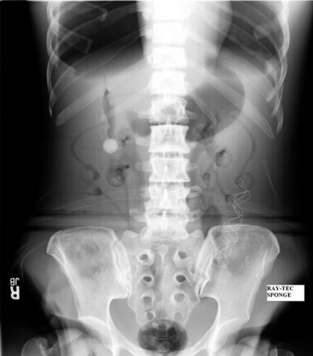
Surgeons typically use two types of sponges in the OR: Ray-Tec sponges and standard lap pads. Ray-Tecs look like a 4×8 piece of gauze with a mysterious blue string woven throughout it. The string is the only part that shows up on x-ray, and it is very thin and somewhat hard to see. Here are some Ray-Tec sponges outside the body:

And here’s one that was left inside. Note the little squiggle in the left lower quadrant and how easy it is to overlook.

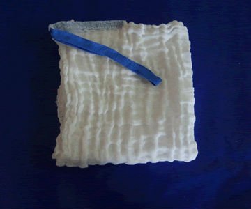
On the other hand, a laparotomy pad is a 4×4 folded cloth pad that unfolds into a larger pad. It has a blue radiopaque tag sewn in the corner, extending along one edge of the pad. Here’s what they look like outside the body:

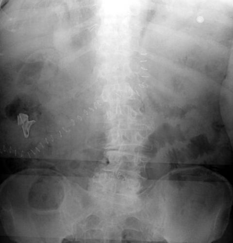
And here’s one inside a patient. Note the irregular object in the right upper quadrant. Many times the tag is scrunched up and doesn’t look like one.

Bottom line: It’s important for anyone who works in the OR on any body part to be familiar with the appearance of these tags on x-rays. Since it’s generally impossible to get accurate counts before or after a trauma procedure, always image the involved body cavity looking for these telltale signs before closing the patient.

Note: These images  were taken from the internet. Patients were not treated at Regions Hospital.