The Lowly Blood Pressure Cuff: Is It Accurate?

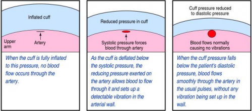
Yesterday, I described how the typical automated oscillometric blood pressure cuff works. We rely on this workhorse piece of equipment for nearly all pressure determinations outside of the intensive care unit. So the obvious question is, “is it accurate?”

Interestingly, there are not very many good papers that have ever looked at this! However, this simple question was addressed by a group at Harvard back in 2013. This study utilized an extensive ICU database from 7 ICUs at the Beth Israel Deaconess Medical Center. Seven years of data were analyzed, including minute by minute blood pressure readings in patients with both automated cuffs and indwelling arterial lines. Arterial line pressures were considered to be the “gold standard.”

Here are the factoids:

  • Over 27,000 pairs of simultaneously recorded cuff and arterial line measurements from 852 patients were analyzed
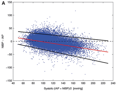
  • The cuff underestimated art line SBP for pressures at or above 95 torr
  • The cuff overestimated SBO for pressures below 95 torr (!)
  • Patients in profound shock (SBP < 60) had a cuff reading 10 torr higher
  • Mean arterial pressure was reasonably accurate in hypotensive patients

sbp-cuff-v-aline

Bottom line: The good, old-fashioned automated blood pressure cuff is fine for patients with normal pressures or better. In fact, it tends to understimate the SBP the higher it is, which is fine. However, it overestimates the SBP in hypotensive patients. This can be dangerous! 

You may look at that SBP of 90 and say to yourself, “that’s not too bad.” But really it might be 80. Would that change your mind? Don’t get suckered into thinking that this mainstay of medical care is perfect! And consider peeking at the mean arterial pressure from time to time. That may give you a more accurate picture of where the patient really is from a pressure standpoint.

Related posts:

Reference: Methods of blood pressure measurement in the ICU. Crit Care Med Journal, 41(1): 34-40, 2013.


How Does It Work? The Lowly Blood Pressure Cuff

The blood pressure cuff is one of those devices trauma professionals don’t give a second thought to. Old timers like me remember using the cuff with a sphygmomanometer and stethoscope to get manual blood pressures. I’ve had to do this twice in recent months on airplanes, and I had forgotten how much work this is.

But technology makes things easier for us. Now you just slap a cuff on the arm (or wrist), push a button, and voila! You’ve got the pressure.

But have you stopped to think about how this actually works? Why don’t we need the stethoscope any more? Here’s the scoop:

When you take a manual blood pressure, the cuff is inflated until a pulse can no longer be auscultated with the stethoscope. The pressure is slowly released using a little thumb wheel while listening for the pulse again. The pressure at which it is first audible is the systolic, and the pressure at which it softens and fades away is the diastolic.

The automatic blood pressure device consists of a cuff, tubing that connects it to the monitor, a pressure transducer in line with the tubing, a mini air pump, and a small computer. The transducer replaces the analog pressure gauge, and the pump and computer replace the human.

The transducer can “see” through the tubing and into the cuff. It is very sensitive to pressure and pressure changes. The computer directs the pump to inflate to about 20 torr above the point where pulsations in the air column cease. It then releases the pressure at about 4 torr per second, “feeling” for air column vibrations to start. When this occurs, the systolic pressure is recorded. Deflation continues until the vibrations stop, representing the diastolic pressure.

bpcuff

Piece of cake! But here’s the question: is it accurate? Tomorrow, I’ll write about how the automated cuff compares to an indwelling arterial line.

Related post:

How Many Salt Tabs In A Liter Of Saline?

Seems like a simple, silly question, right? I dare you to figure it out without reading this post!

horse-salt-block-lick2

On occasion, our brain injured trauma patients have sodium issues. You know, cerebral salt wasting. Trying to maintain or regain the normal range, without making any sudden moves can be challenging. There are a lot of tools available to the trauma professional, including:

  • Saline
  • Hypertonic saline
  • Salt tablets
  • Fluid restriction
  • Some combination thereof

Fun times are had trying to figure out how much extra sodium we are giving with any of the first three items. This is important as you begin to transition from the big guns (hypertonic), to regular saline, and then to oral salt tabs.

Below is a quick and dirty conversion list. I won’t make your heads explode by trying to explain the math involved changing between meq, mg, moles, sodium and sodium chloride.

  • The “normal saline” bags we use are actually 0.9% saline (9 gm NaCl per liter)
  • Hypertonic saline can be 3% or 5% (30 gm or 50 gm per liter)
  • Salt tabs are usually 1 gm each (and oh so yummy)

Therefore, a liter of 0.9% normal saline is the same as 9 salt tabs.

A liter of 3% hypertonic saline is the same as 30 salt tabs. The usual 500cc bag contains 15.

A liter of  5% hypertonic saline is the same as 50 salt tabs. The usual 500cc bag contains 30.

To figure out how many tablets you need to give to match their IV input, calculate the number of liters infused, then do the math! And have fun!

Next Trauma MedEd Newsletter Is Coming Next Week!

As promised, the next Trauma MedEd newsletter will be released next week. Just in time for some light Christmas reading!

The topic is “Prevention.” Here are the areas I’ll be covering:

  • The American College of Surgeons requires all US trauma centers to engage in prevention activities. Unfortunately, there is frequently confusion about the role of the injury prevention coordinator, what kinds of programs are acceptable, and how local data needs to be included in prevention planning. I will cover all of this, and more, in the first part of the newsletter.
  • Curious about what others are doing out there? I’ll give you an idea of the most common prevention programs, and whether they are national programs or home grown.
  • I’ll review a few papers on the efficacy of trauma prevention programs.
  • Finally, I’ll give some tips on how to optimize the performance of your injury prevention coordinator and design effective programs.

As always, this issue will go to all of my subscribers first. If you are not yet one of them, click this link to sign up and/or download back issues.

Unfortunately, non-subscribers will have to wait until I release the issue on this blog, sometime during the week after Christmas. So sign up now!

The IVC Filter In Trauma: Why?

The inferior vena cava (IVC) filter has been around in one form or another for over 40 years. One would think that we would have figured everything about it out by now. But no!  The filter has evolved through a number of iterations and form factors over the years. The existing studies, in general, give us piecemeal information on the utility and safety of the device.

One of the major innovations with this technology came with the development of a removable filter. Take a look at the product below. Note the hook at the top and the (relatively) blunt tips of the feet. This allows a metal sheath to be slipped over the filter while in place in the IVC. The legs collapse, and the entire thing can be removed via the internal jugular vein.

ivc-filter-complications1

The availability of the removable filter led the American College of Chest Physicians to recommend their placement in patients with known pulmonary embolism (PE) or proximal deep venous thrombosis (DVT) in patients with contraindications to anticoagulation. Unfortunately, this has been generalized by some trauma professionals over the years to include any trauma patients at high risk for DVT or PE, but who don’t actually have them yet.

One would think that, given the appearance of one of these filters, they would be protective and clots would get caught up in the legs and be unable to travel to the lungs as a PE. Previous studies have taught us that this is not necessarily the case. Plus, the filter can’t stop clots that originate in the upper extremities from becoming an embolism. And there are quite a few papers that have demonstrated the short- and long-term complications, including clot at and below the filter as well as post-phlebitic syndrome in the lower extremities.

A new study from Boston University reviewed their own experience retrospectively over a 9 year period. This cohort study looked at patients with and without filters, matching them for age, sex, race, and injury severity. The authors specifically looked at mortality, and used four study periods during the 9 year interval.

Here are the factoids:

  • Over 18,000 patients were admitted during the study period, resulting in 451 with an IVC filter inserted and 1343 matched controls
  • The patients were followed for an average of 4 years after hospitalization
  • Mortality was identical between patients with filters vs the matched controls

dvt-study

  • There was still no difference in mortality, even if the patients with the filter had DVT or PE present when it was inserted
  • Only 8% ever had their “removable” filter removed (!)

Bottom line: Hopefully, it’s becoming obvious to all that the era of the IVC filter has come and gone. There are many studies that show the downside of placement. And there are several (including this one) that show how forgetful we are about taking them out when no longer needed. And, of course, they are expensive. But the final straw is that they do not seem to protect our patients like we thought (hoped?) they would. It’s time to reconsider those DVT/PE protocols and think really hard about whether we should be inserting IVC filters in trauma patients at all.

Related post:

Reference: Association Between Inferior Vena Cava Filter Insertion
in Trauma Patients and In-Hospital and Overall Mortality. JAMA Surg, online ahead of print, September 28, 2016.