Field Amputation: Question for Readers

I’m going to be revisiting my series on field amputation over the next few days. We will be reviewing our own policies and procedures regarding this topic at the trauma operations committee here at Regions Hospital. 

I’d like to query my readers, all of you trauma professionals out there in any discipline. Who is qualified to perform this procedure? What special training is required? Can it safely be done by an emergency physician? A paramedic? A nurse? I’ve reviewed quite a bit of the literature. But as we all know, research and reality don’t always coincide. And I’m particularly interested in responses from my international readers.

Please take a moment to email or leave comments below. I’ll compile them over the next few days and publish a compendium next week. Thanks!

Antiplatelet Therapy And Blunt Head Trauma

All trauma professionals are aware of the evils of anticoagulation in patients who sustain traumatic brain injury. Warfarin is one of the most common anticoagulants encountered, but there is also a growing number of poor outcomes in patients with the newer, non-reversible agents.

But what about antiplatelet agents like aspirin and clopidogrel (Plavix)? Many physicians worry about these drugs, but is it warranted? Two Level I trauma centers in the Chicago area reviewed their experience. They retrospectively reviewed the records of patients over 40 years old who sustained blunt head trauma. A total of 1547 patients were identified over a 4 year period. They analyzed these records for in-hospital mortality, need for neurosurgical intervention, and length of stay.

Here are the factoids:

  • 27% of patients were taking antiplatelet agents. Patients also taking warfarin were excluded.
  • 21% were taking aspirin alone, 2% clopidogrel alone, and 4% both drugs
  • Patients taking the drugs averaged about 10 years older than those who were not
  • Overall, injury severity was relatively low (average ISS 10). A disproportionate number of more severely injured patients were not taking antiplatelet agents.
  • There was no difference of incidence of intracranial hemorrhage (45%), neurosurgical intervention (3%), or mortality (6%) between the two groups
  • Hospital length of stay averaged about 6.5 days, but long LOS was a bit more common in the antiplatelet agent group.

Bottom line: This is one more in a series of papers scrutinizing trauma and antiplatelet agents. A few previous studies have shown an adverse effect, but they have been much smaller series. I don’t believe the jury is in yet, so watch these patients carefully. A 6 or 12 hours repeat scan is probably in order, along with frequent neuro monitoring.  It’s probably not worthwhile to actively try to reverse them by giving platelets unless there is obvious life-threatening hemorrhage or sudden neurologic change (see below).

Related posts:

Reference: Outcomes in traumatic brain injury for patients presenting on antiplatelet therapy. Am Surg 81(2):128-132, 2015.

Another Failure Of Shotgun Style Diagnostic Testing: The Trauma Incidentaloma

When our patients present with a problem, there is a time honored and well-defined sequence to help us come to a final diagnosis. 

  • Take a detailed history
  • Examine the patient
  • Order pertinent diagnostic tests, if indicated
  • Then think about it a while

The first two items are a chip shot, and the trauma professional can gain a lot of information by spending a relatively short period of time doing these. And many times the diagnosis can be made without any further action.

However, diagnostic testing of all kinds has become so prevalent and easy to obtain that we rely on it a bit too much. And sometimes, we order it up in lieu of a thorough history and exam. If the clinician skimps on those steps, it’s much more difficult to narrow the list of differential diagnoses to a manageable number.

So what happens then? They use diagnostic tests as a crutch. Instead of being able to select a few focused tests to answer the questions, they essentially put an order sheet on the wall, fire off a shotgun, and order everything that’s been hit by the pellets. 

Lots of tests, so they will definitely find the answer, right? Nope! There are two major problems here. First, the so-called signal to noise ratio is very low. There are so many results, that it is easy to overlook a pertinent positive among all the negatives.

But more significantly, there is always the possibility that there will be more than one positive. One of them might actually be the answer you were seeking. But what about the others? There are the trauma incidentalomas. Some may be truly positive, but there is always the possibility of a false positive. These are the most treacherous, because many trauma professionals then feel obligated to “do something about it.” 

As we have found from multiple screening tests like PSA, PAP smear, and mammography, a significant number of patients may be harmed trying to further investigate what turns out to be nothing at all (artifact), or something completely benign. This includes not only harm from complications or unnecessary procedures, but months of anxiety the patient may suffer while the clinicians figure out what that thing inside them really is.

There are only a few studies on trauma incidentalomas available. One reviewed a series of almost 600 head CT scans in patients with TBI and found unexpected findings on 85%. About 90% were obviously benign. Unfortunately, it was not possible to follow these patients to find out how many of the remaining lesions turned out to be benign as well. But I would wager that most did.

Bottom line: I shouldn’t even have to say this, but do a good history and physical exam! If you need diagnostic studies, order only the one(s) that have the potential to make your final diagnosis. Don’t shotgun it. One very helpful tool is a well-designed practice guideline for commonly encountered clinical scenarios. This will limit the number of “other” findings you have to deal with. And finally, did I say to do a good history and physical exam?

Related posts:

Reference: Incidental cranial CT findings in head injury patients in a Nigerian tertiary hospital. J Emerg Trauma Shock 8(2):77-82, 2015.

April Trauma MedEd Newsletter Released

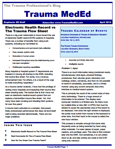
The April newsletter is now available! Click the image below or the link at the bottom to download. This month’s topic is “ED Stuff”.

In this issue you’ll find articles on:

  • Lots of info on trying to use an electronic trauma flow sheet
  • How do you dress your trauma team?

Subscribers received the newsletter earlier this week. If you want to subscribe to get early delivery in the future (and download back issues), click here.

Click here to download.

Liver Laceration And Liver Function Tests

Over the years I’ve seen a number of trauma professionals, both surgeons and emergency physicians, order liver transaminases (SGOT, SGPT) and bilirubin in patients with liver laceration. I’ve never been clear on why, so I decided to check it out. As it turns out, this is another one of those “old habits die hard” phenomena.

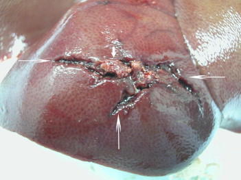
Liver lacerations, by definition, are disruptions of the liver parenchyma. Liver tissue and bile ducts of various size are both injured. Is it reasonable to expect that liver function tests would be elevated? A review of the literature follows the typical pattern. Old studies with very few patients.

From personal hands-on observations, the liver tissue itself tears easily, but the ducts are a lot tougher. It is fairly common to see small, intact ducts bridging small tears in the substance of the liver. However, larger injuries can certainly disrupt major ducts, leading to major problems. But I’ve never seen obstructive problems develop from this injury.

A number of papers (very small, retrospective series) have shown that transaminases can rise with liver laceration. However, they do not rise reliably enough to be a good predictor of either having an injury, or the degree of injury. Similarly, bilirubin can be elevated, but usually not as a direct result of the injury. The most common causes are breakdown of transfused or extravasated blood, or from critical care issues like sepsis, infection, and shock.

Bottom line: Don’t bother to get liver function tests in patients with known or suspected injury. Only a CT scan can help you find and/or grade the injury. And never blame an elevated bilirubin on the injury. Start searching for other causes, because they will end up being much more clinically significant.

References:

  • Evaluation of liver function tests in screening for intra-abdominal injuries. Ann Emerg Med 20(8):838-841, 1991.
  • Markers for occult liver injury in cases of physical abuse in children. Pediatrics 89(2):274-278.
  • Combination of white blood cell count with liver enzymes in the diagnosis of blunt liver laceration. Am J Emerg Med 28(9):1024-1029, 2010.