So yesterday, we found that our patient was hemodynamically stable, with a knife in his back, positioned prone. An initial chest xray shows the knife (plainly) and haze in the right side of the chest. Obviously, this is a hemothorax.
Key points to note are that the amount of blood present is modest and the knife point is relatively medial, as is the entry seen on the outside. Combined, these data points indicate that you have time to gather more information.
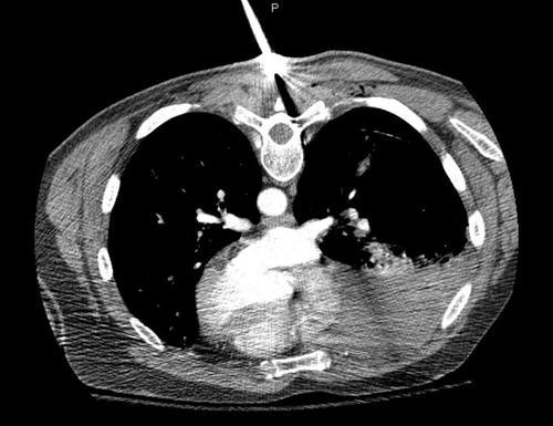
My choice was to go to CT to get the ultimate anatomic information. What, you say, the patient is prone! Well, the scanner doesn’t care. As long has his torso AND the knife fit through, it works. Here’s the representative scan result:
What do you do now? Where do you do it? Answer tomorrow. Tweet or comment your decision!
Related posts:
