EAST 2014: Spine Imaging In Elderly Falls

Many people don’t realize, but falls are more common than motor vehicle crashes. The elderly are most commonly involved, and injuries frequently have a major impact on quality of life. Dogma tells us that we should image the full spine if any part of it is fractured. 

A group at Thomas Jefferson University questioned this practice (good!) and designed a study to look at its efficacy. They hypothesized that the low energy involved would not cause enough non-contiguous spine injuries to be of concern.

They designed a retrospective study using a large pool of data from the Pennsylvania Trauma Systems Foundation trauma registry. Here are the factoids:

  • Only patients older than 65 who fell from standing and sustained a cervical spine fracture were included
  • Of over 14,500 elderly patients who fell, 1102 sustained a cervical fracture
  • 1083 of these patients were neurologically intact (99%) and the status of the remainder of their spine was evaluated
  • 7% of neuro intact patients with a cervical fracture also had a thoracolumbar spine fracture
  • Three of these 74 patients required a surgical spine procedure
  • The presence of a rib fracture was associated with triple the incidence of a thoracolumbar fracture

Bottom line: Although this study looks convincing, there are a few issues. First, it’s a registry study and data quality is always a concern. This may explain the lower than usual incidence of thoracolumbar fractures after fall from standing compared to other reports. And based on their work, the authors recommend CT screening of the T and L spines if a cervical fracture is present. This may be overkill, and an initial screen with conventional spine xrays may help decrease the number of spine CTs performed, even though sensitivity and specificity for these studies is low.

Reference: Is full spine imaging necessary in the elderly, fall from standing trauma patient with a cervical fracture? EAST 2014, oral paper 13.

Related post:

The December issue of Trauma MedEd is ready! Subscribers will receive it New Years Eve! Perfect to have a few drinks over! This issue is devoted to trauma PI, which brings tears to many an eye

Included are articles on:

  • System vs peer issues
  • Loop closure
  • Over- and undertriage
  • Trauma filters
  • And more!

As mentioned above, subscribers will get the issue delivered New Year’s Eve to their preferred email address. It will be available to everybody else next week on the blog.

Check out back issues, and subscribe now! Get it first by clicking here!

EAST 2014: The Flat Vena Cava Sign?

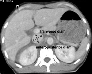
I’ve previously blogged about the flat vena cava sign as an indicator of low volume status in trauma patients. One of the papers at EAST takes another look at this tool, and had a surprisingly negative result.

A retrospective study at George Washington University was carried out over a one year period. They looked at all of their highest level trauma activation patients who also underwent CT scan of the abdomen. Images were read by three radiologists and inter-rater reliability was reviewed. The transverse to anteroposterior diameter ratios were calculated to determine flatness.

Here are the factoids:

  • 276 patients met enrollment criteria, and were mostly male and blunt trauma
  • The IVC was nearly round in 21% of patients and collapsed in 26%
  • There was no association between IVC shape and shock index, blood pressure, Hbg, lactate, urgent operation, angiography or length of stay
  • There was also no association between IVC shape and blood transfusion or death
  • Correlation of the reads between radiologists was good

So what gives? A paper I reviewed three years ago in the Journal of Trauma came to a different conclusion. They found that a flat IVC on CT scan (defined as a transverse to AP ratio of 4:1 or greater) was associated with a significantly higher chance of receiving more crystalloid or blood, as well as requiring an operation within 24 hours. 

First, the newer work is an abstract, so a lot may be unsaid at this time. This is why I encourage everyone to always read the entire paper! The published paper involved a smaller series (114 patients), but it was prospective and had reasonable statistical analyses.

Bottom line: This is a presentation that I’ll have to sit through and ask the authors why they didn’t find the same results as the older paper. For now, continue to use the flat IVC sign as a potential sign of trouble ahead. I’ll report more on this one later in January.

Reference:

  • Inferior vena cava size is not associated with shock following injury. EAST 2014, oral paper 12.

Related post:

The Best (And Worst) Of EAST

The EAST meeting is upon us in just a few weeks! I’ll be attending (and tweeting) during this annual event. But in the meantime, I’d like to provide some commentary on some of the best and worst abstracts that will be presented.

Starting tomorrow, I’ll summarize one interesting paper/poster per day. I’ll also provide some perspective as to why it looks important (or not). Remember, my impressions are based on just an abstract. There is a lot that is left unsaid in the preparation of these brief research summaries. Sometimes, those things are crucial and totally destroy the work promised in the abstract.

I’ll also be listening to the presentations of the papers I critique at the meeting so I can get the real story, and I’ll share those with you as well next month.