The 8 Hour Rule For Open Fractures: We’re So Over That

For decades, the standard of care for irrigation and debridement (I&D) of open fractures has been within 8 hours of injury. There is a growing body of orthopedic literature that says this isn’t necessarily so.

A paper being presented at the AAST meeting in Chicago next week retrospectively looked at their experience with early (<8hrs) vs late I&D in a series of 248 patients. They looked at infection rates stratified by time and upper vs lower extremity.

They found that the infection rates overall were not significantly different. However, when subgrouped by extremity and higher Gustilo type >= III, they noted that both delayed I&D and Gustilo type correlated with infection risk. For the upper extremity, only Gustilo type >= III correlated with a higher infection rate.

The authors concluded that all lower extremity open fractures should be dealt with in the 8 hour time frame, whereas upper extremity fractures can be delayed for lower Gustilo classes.

Bottom line: I don’t necessarily buy into all the results from this small study. The orthopedic literature has already refined this concept. At Regions Hospital, we allow up to 16 hours to I&D for open fractures up to and including Gustilo class IIIA. Above that, the 8 hour rule is followed. We periodically review our registry data on all open fracture patients to make sure that the extended time frame patients are not experiencing an increase in wound complications. And they haven’t in our 8 year experience in handling them this way.

Refresher on the Gustilo classification system:

  • Class I – open fracture, clean wound, <1cm laceration
  • Class II – clean wound, laceration >1cm with minimal soft tissue damage
  • Class IIIA – clean wound, more extensive soft tissue damage or laceration, periosteum intact, minimal contamination
  • Class IIIB – extensive soft tissue damage with periosteal stripping or bone damage, significant contamination
  • Class IIIC – arterial injury without regard for degree soft tissue injury

Reference: Open extremity fractures: does delay in operative debridement and irrigation impact infection rates? AAST 2011 Annual Meeting, Paper 22.

What To Do? Small Hospital, Unstable Patient

It’s the situation that physicians in smaller hospitals dread. A major trauma patient gets dropped off at the door. You do your evaluation, quickly determining that they need services that you don’t just have (head injury and positive FAST in the abdomen, let’s say). You call your community EMS service to transport to a Level I trauma center, which is about 30 minutes away by ground. And just as they are rolling out the door to the rig, the blood pressure drops to 60! What to do?!

The ATLS course is very clear, and very correct. Back into the ED for a quick re-evaluation. The most common cause for a significant disturbance in vitals or exam lies within the primary survey. You will almost always find a problem with Airway, Breathing, or Circulation. (A Disability problem can cause a problem on rare occasion (hypotension from impending herniation), but there’s not much that you can do about it, really. Hyperventilate, hyperosmolar therapy, okay but probably a poor outcome for the patient anyway.)

So you didn’t find any airway or breathing issue. But the abdominal stripe(s) you saw on FAST are larger, so it’s circulation. Now what? And does it matter if you have a surgeon available on call? The answer is simpler than you think.

ATLS says that, if you have surgical support available you have to use it in this type of situation. If you don’t have it, package the patient with a lot of blood and plasma and send. If you have a physician or nurse to spare you could consider sending them along to help during transport, but for small community hospitals this is not practical.

But if you do have a surgeon, does it make sense to use them? Not always! You must take into account response times and transport times. Let’s say it’s 2:00 am and you call your surgeon for this hypotensive patient. They may take up to 30 minutes to get in and see the patient. They then agree that the patient needs a laparotomy and she proceeds to call in the OR team. Yet another 30 minutes tick by.  Will the patient still even be alive when they roll into the OR?

Or you could just put the patient back in the ambulance (air preferably, but ground if you have to) and get them to your trauma center quickly. They can then be whisked directly into a waiting OR in less than 30 minutes from your door. This is probably the ideal solution here. Obviously this doesn’t work as well if you are a few hours away from your resource trauma center. 

Bottom line: Deciding what to do with a patient that needs urgent treatment that you can’t immediately deliver is tough! That’s why it’s always a calculus problem when you’re faced with this situation. But take all of the response and transport times into account, and do what’s best for your patient! 

Thanks to EM Res for posing this question!

Above-Knee vs Below-Knee DVT

Deep venous thrombosis (DVT) is a common problem in trauma patients. Many trauma centers have developed practice guidelines for beginning mechanical or chemoprophylactic measures as soon as practical in patients at risk. Some believe that below-knee and above-knee DVT are different, with those located below the knee posing a lower risk for propagation and pulmonary embolism. As always, we need to know, is it true?

The group at Oregon Health and Science University performed a retrospective review of six years of their experience with lower extremity DVT. They identified 308 patients who developed this complication and noted the following interesting findings:

  • Two thirds developed below-knee DVT, one third above-knee
  • Overall rate of pulmonary embolism (PE) was about 4% overall
  • PE occurred with equal frequency in below-knee vs above-knee DVT
  • The rate at which DVT resolved was no different in patients receiving prophylactic doses of enoxaparin vs therapeutic dosing
  • Below-knee DVT did not resolve faster than above-knee. Thus they are not more likely to resolve spontaneously

The point of looking for and giving enoxaparin and similar drugs in trauma patients is to avoid DVT, limit it’s progression, and prevent PE. This study showed that there really is no difference between below-knee and above-knee DVT, and that they should be treated similarly. Unfortunately, it also showed that prophylactic and therapeutic management worked equally as well. This is probably due to the fact that there are major differences across various types of trauma patients and that we still don’t know how to calculate the right dose of enoxaparin. However, we do have some tools to help us make a better guess. 

Bottom line: Trauma patients with any lower extremity DVT need to be treated, and enoxaparin is a common way to do this. Below-knee vs above-knee does not matter. If enoxaparin is used, just selecting a therapeutic dose (e.g. 1mg/kg bid) is not enough. Monitoring with anti-factor Xa levels or thromboelastogram (TEG) may help optimize effectiveness and reduce risk of PE.

Related posts:

Reference: The effects of location and low-molecular-weight heparin administration on deep vein thrombosis outcomes in trauma patients. J Trauma 74(2):476-481, 2013.

January TraumaMedEd Newsletter

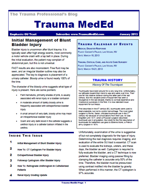
The January newsletter is here! Click the image below or the link at the bottom to download. This month’s topic is Genitourinary, providing information on:

  • Initial management of bladder injury
  • How to do CT cystogram
  • Extraperitoneal bladder injury
  • Followup cystogram
  • Retrograde urethrogram in patients with a catheter in place
  • Renal injury grading update

Subscribers had the newsletter emailed to them on Tuesday. If you want to subscribe (and download back issues), click here.

Download the newsletter

The Two-Sheet Trauma Trick

Hypothermia is always a concern in trauma patients. Even the simple act of completely exposing your patient in the trauma room facilitates it. How do trauma professionals balance the need to see everything with the equally important need to keep the patient warm?

The natural reaction is to cover them up. Sheets and warm blankets are the usual tools. But I always marvel that, as soon as the blanket goes on, there’s always a need to examine something or do some procedure. Look at a wound. Insert a urinary catheter. And every time this happens, the blanket comes off.

Here’s a clever way to deal with this problem. Don’t use just one sheet or blanket. Use two! Fold each one in half, so they are each half-length. Place one on the top half of the patient, the other at the bottom, overlapping slightly at the waist. If you need to look at an extremity, fold the blanket that covers it over from right to left (or left to right) to uncover just the area of interest. To insert a urinary catheter, just open the area at the waist, moving the top sheet up a little, the bottom down a little.

Bottom line: Keep your patient toasty! Use the two-sheet (or warm blanket) trick to avoid hypothermia. Remember, patient temperature begins to drop as soon as the clothes come off! And I don’t recommend the use of one-piece inflatable warming blankets (e.g. Bair Hugger) until the work in the ED is complete, because the whole thing has to be removed every time you need meaningful access to the patient.

Related posts: