Pop Quiz: Hints

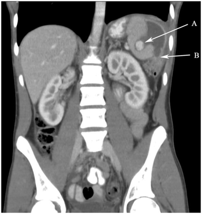
No correct guesses yet, but some good tries! Remember, this was a 16 year old male who crashed a motorcycle and sustained liver and spleen injuries. A month after successful nonoperative management, he presents with a single episode of hematochezia. 

Here’s a CT scan taken during the second ED visit:

What’s the problem? Any way to have detected it sooner? What to do now?

Comment or tweet your answers!

Pop Quiz: Interesting Case!

A 16 year old male was thrown against the handlebars during a motorcycle crash at about 40 mph. He dusted himself off and went home for a few hours. Unfortunately, he slowly developed some abdominal pain.

He presented to an ED several hours later. He was found to have mild, diffuse abdominal pain, normal vital signs, and a positive abdominal FAST exam. CT scan showed a grade IV spleen injury and a grade II liver injury in the right lobe with no extravasation or pseudoaneurysm noted. He was successfully treated nonoperatively and was sent home.

One month later he returns to the ED complaining of a single episode of hematochezia (approximately 200cc). He has an entirely normal exam and vital signs.

Here are my questions for you:

  • Was the initial management appropriate?
  • Should anything additional have been done during the first admission?
  • What is the diagnosis now?
  • What diagnostic or therapeutic maneuvers are indicated now?

Please tweet your guesses, or leave comments below. Hints tomorrow and answers on Friday. Good luck!

Patient not treated at Regions Hospital

What’s The INR Of Fresh Frozen Plasma?

So what’s the INR of FFP? Or stated another way, what’s the lowest you can correct a patient’s INR using infusions of fresh frozen plasma?

One of the mainstays of correcting coagulopathy, either from hemorrhage or due to medication like warfarin, is transfusion of FFP. Frequently, clinicians will write orders to administer FFP until a certain INR is achieved. What is a reaonable INR?

A “normal” INR is 1.0, plus or minus about 0.2, depending on your laboratory. However, two separate studies have shown that transfusion of FFP will not reliably decrease the INR below about 1.7. 

Bottom line: The answer to the question is about 1.6. If any clinician orders FFP transfusions with a goal INR below this, it probably won’t happen. And since transfusions of any product have risks, my “juice to squeeze” ratio of risk vs benefit begins to fail at an INR of 1.6. Below that point, the patient needs a normal temperature and good perfusion to drop their INR further.

References:

  • Toward rational fresh frozen plasma transfusion: the effect of plasma transfusion on coagulation test results. Am J Clin Pathol 126(1):133-139, 2006.
  • Effect of fresh frozen plasma transfusion on prothrombin time and bleeding in patients with mild coagulation abnormalities. Transfusion 46(8):1279-1285, 2006.

Best Of: CT Contrast Via Intraosseous Catheter

The standard of care in vascular access in trauma patients is the intravenous route. Unfortunately, not all patients have veins that can be quickly accessed by prehospital providers. Introduction of the intraosseous device (IO) has made vascular access in the field much more achievable. And it appears that most fluids and medications can be administered via this route. But what about iodinated contrast agents via IO for CT scanning?

Physicians at Henry Ford Hospital in Detroit published a case report on the use of this route for contrast administration. They treated a pedestrian struck by a car with a lack of IV access sites by IO insertion in the proximal humerus, which took about 30 seconds. They then intubated using rapid sequence induction, with drugs injected through the IO device. They performed full CT scanning using contrast injected through the site using a power injector. Images were excellent, and ultimately the patient received an internal jugular catheter using ultrasound. The IO line was then discontinued.

This paper suggests that the IO line can be used as access for injection of CT contrast if no IV sites are available. Although it is a single human case, a fair amount of studies have been done on animals (goats?). The animal studies show that power injection works adequately with excellent flow rates. 

The authors prefer using an IO placement site in the proximal humerus. This does seem to cause a bit more pain, and takes a little practice. A small xylocaine flush can be administered to reduce injection discomfort in awake patients. Additionally, the arm cannot be raised over the head for the torso portion of the scan. 

Bottom line: CT contrast can be injected into an intraosseous line (IO) with excellent imaging results. Insert the IO in a site that you are comfortable with. I do not recommend power injection at this time. Although the marrow cavity can support it, the connecting tubing may not. Have your radiologist hand-inject and time the scan accordingly.

Note: long term effects of iodinated contrast in the bone marrow are not known. For this reason, and because of smaller marrow cavities, this technique is not suitable for pediatric patients.

Related posts: 

Reference: Intraosseous injection of iodinated computed tomography contrast agent in an adult blunt trauma patient. Annals Emerg Med 57(4):382-386, 2011.
Orthopedic Hardware And TSA Metal Detectors

Many trauma patients require implantable hardware for treatment of their orthopedic injuries. One of the concerns they frequently raise is whether this will cause a problem at TSA airport screening checkpoints (Transportation Safety Administration)

The answer is probably “yes.” About half of implants will trigger the metal detectors, and these days that usually means a pat down search. And letters from the doctor don’t help. It turns out that overall, 38% are detected when the scanner is set to low sensitivity and 52% at high sensitivity. 

Here is a more detailed breakdown:

  • Lower extremity hardware is detected 10 times more often than upper extremity or spine implants
  • 90% of total knee and total hip replacements are detected
  • Upper extremity implants such as shoulder, wrist and radial head replacements are rarely detected
  • Plates, screws, IM nails, and wires usually escape detection
  • Cobalt-chromium and titanium implants trigger alarms more often than stainless steel

 If your patient knows that their implant triggers the detectors, they have two options: request a patdown search, or volunteer to go through the full body millimeter wave scanner. This device looks at everything from the skin outwards, and will not “see” the implant and is probably the preferred choice. If they choose to go through the metal detector and trigger it, they are required to have a patdown. Choosing to go through the body scanner after setting off the detector is no longer an allowed option. 

Reference: Detection of orthopaedic implants in vivo by enhanced-sensitivity, walk-through metal detectors. J Bone Joint Surg Am. 2007 Apr;89(4):742-6.