Spleen Week

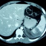
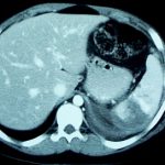
This week I’ll be covering spleen injuries. The answer to the question “What is wrong with this spleen” is: 1. There is a spleen laceration (grade cannot be determined from this one slice) and 2. There is a contrast blush.

Today I’ll cover grading and tomorrow I’ll talk about the significance of blushes.

How Often Are Imaging Studies Repeated After Trauma Transfers?

Smaller trauma hospitals, both designated and undesignated, are the front line for the initial care of the majority of trauma patients. Many patients can be evaluated and sent home or admitted to the initial hospital. More severely injured patients are commonly transferred to the nearest Level I or Level II trauma center for care of injuries requiring specialists.

Imaging studies such as conventional xray and CT scan are a necessary part of the initial trauma evaluation. But is it necessary to do a full radiographic evaluation, even when it is known that the patient will have to be transferred?

Researchers at Dartmouth Hitchcock Medical Center examined the issue of repeat imaging at their Level I center. They looked at 138 patients that were transferred to them from other rural hospitals. They found that 75% underwent CT scanning prior to transfer, and 58% underwent repeat scanning upon arriving at Dartmouth.

The authors discovered the following:

  • Head CTs were repeated 52% of the time, primarily due to clinical indications
  • Spine reconstructions were repeated 33-50% of the time due to inadequate reconstruction technique
  • Chest (31%) and abdomen (20%) were repeated due to inappropriate use of IV contrast
  • 13% of image disks used incompatible software
  • 7% of images were not sent with the patient

Here are my recommendations for imaging by hospitals that refer patients to Level I or II trauma center:

  • Obtain the essential plain films recommended by ATLS (chest, pelvis)
  • If an obvious injury requiring transfer is found on exam (e.g. open fracture) do no further studies
  • Obtain any imaging studies needed to decide if you can admit the patient to your own hospital (example: abdominal CT for abdominal pain and negative FAST. Keep if no injury, transfer if solid organ injury)
  • As soon as an injury is identified that mandates transfer, do no further studies
  • Always send image disks with the patient
  • Work with your referral trauma center to obtain a copy of their CT imaging protocols so if you do need to perform a study you can duplicate their technique

Reference: Gupta et al. Inefficiencies in a Rural Trauma System: The Burden of Repeat Imaging in Interfacility Transfers. J Trauma 69(2):253-255, 2010.

Top 10 Worst Complications: #1 Nasocerebral Tube

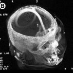
Minor complications from nasogastric tube insertion occur relatively frequently. Emesis is fairly common when the gag reflex is stimulated by the tube in the back of the oropharynx. An infrequent but possibly fatal one is insertion through the cribriform plate. 

The cribriform plate is located directly posterior to the nares and is part of the ethmoid bone. It is very porous in nature and weaker than the surrounding portions of the ethmoid. It is easily fractured, and can be seen is association with basilar skull fractures. This is one source for rhinorrhea in patients with these fractures.

Cribriform fracture is a contraindication to unprotected insertion of a nasogastric tube. If you look at the sagittal section below, the plate lies directly behind the nares. When inserting the NG tube, we are usually taught to aim the tube straight back. Unfortunately, this aims it directly at the cribriform. If a fracture is present, it is possible that you may be inserting a nasocerebral tube!

Cribriform plate - sagittal section

The usual symptoms when this occurs consist of immediate neurologic deterioration to coma, and a unilateral or bilateral blown pupil. The tube must not be withdrawn, because it will cause significant injury to the base of the brain. A stat neurosurgical consultation must be obtained, and if the patient is salvageable, the tube must be withdrawn through a craniectomy.

To avoid this dreaded complication, identify patients at risk for cribriform injury. They are:

  • patients with signs of trauma from eyebrows to zygoma
  • comatose patients
  • patients with signs of basilar skull fracture (Battle’s sign, raccoon eyes, oto- or rhinorrhea)

If your patient is at risk, follow these guidelines:

  • first, does the patient really need a gastric tube?
  • if comatose, insert an orogastric tube
  • if awake, don’t put the tube in their mouth, as they will gag continuously. Instead, place a lubricated, curved nasal airway. Then lube up a slightly smaller Salem sump tube and pass it through the airway.