Tag Archives: extravasation

Contrast Extravasation Into The Psoas Muscle

Contrast extravasation after major trauma can be very problematic. Extravasation into a solid organ (liver, spleen) generally requires a quick trip to interventional radiology or the operating room. Bleeding from the bowel mesentery assures an exploratory laparotomy. Gluteal vessel extravasation is best treated with angioembolization.

But what about extravasation from off the beaten path areas like the psoas muscle? This is an uncommon finding on trauma CT, so less is known about the usual clinical course. A group in Okayama Japan performed a 10-year retrospective review of data from their hospital. They reviewed hematoma size, associated injuries, and the relationship to treatment options.

Here are the factoids:

  • 762 contrast CTs were performed due to blunt trauma over the 10 year period (only 76 per year?!)
  • About 15% (117 patients) had either lumbar process fracture or psoas hematoma, and about one quarter had obvious contrast extravasation into the muscle
  • Patients with contrast extravasation were significantly older, had higher ISS, and were more likely to require transfusion
  • There was an association between the number of transverse process fractures and “need for” angioembolization
  • Size of the psoas hematoma was predictive of the need for angioembolization
  • Angioembolization of the psoas was frequently associated with  embolization of the pelvis

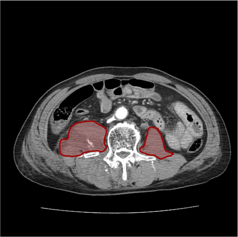
The right psoas has both contrast extravasation and a sizable hematoma

Bottom line: This study has many weaknesses, but does show that psoas extravasation occurs somewhat frequently, even at a low volume center. I always worry about studies that state something like “and xx patients required intervention.” Generally, this means that it was performed at the discretion of the clinician and no consistent rules were applied. And even though hematoma size was significantly correlated with angioembolization, it’s probably not worth the effort to have your radiologist calculate it. But it does illustrate one nearly universal trauma rule:

Patients with active extravasation on CT are bleeding to death until proven otherwise

Do not sit back and manage expectantly! The corollary to this rule is:

Contrast extravasation on CT always requires active measures to stop it

These active measures are typically angioembolization for difficult to reach areas in hemodynamically stable patients (gluteal artery for buttock, lumbar artery for psoas muscle, solid organs). Unstable patients absolutely require a trip to the OR for control. Superficial muscular bleeding frequently stops with good pressure dressings or positioning the patient so they lie on the affected area. Just don’t sit around and watch these patients bleed when you see extravasation on the CT.

Reference: Impact of contrast extravasation on computed tomography of thepsoas major muscle in patients with blunt torso trauma. J Trauma 86(2):268-273, 2019.

Splenic Vascular Blush

Contrast blush is always a concern when seen on CT of the abdomen for trauma. It can represent one of two things, and both are bad:

  • Active extravasation of contrast
  • Splenic pseudoaneurysm

These two clinical issues can be distinguished by looking at the location of the contrast and its persistence. A pseudoaneurysm is located within the parenchyma, and the contrast will wash away, so it will not be visible on delayed images. Contrast that extends beyond the parenchyma or persists in delayed views represents active bleeding. In either case, the failure rate of nonoperative management exceeds 80% in adults without additional measures being taken.

Clinically, these patients usually act as if they are losing volume and require additional crystalloid and/or blood transfusion. The natural history in adults is for bleeding to continue or for the pseudoaneurysm to rupture, resulting in a quick trip to the operating room.

If vital signs can be maintained with fluids and blood, a trip to interventional radiology may solve the problem. Selective or nonselective embolization can be carried out and patients with only a few bleeding points can be spared operation. However, if multiple bleeding areas are seen, it is probably better to head to the OR for splenorrhaphy or splenectomy.

The image below shows likely areas of extravasation. They are a bit large to be pseudoaneurysms.

Spleen Blush-CT

Children are different than adults. Extravasation from spleen injuries in prepubescent children frequently stops on its own. Angiography should only be used if the child is failing nonoperative management.

Next post: A new paper looks at the natural history of these lesions.

Splenic Vascular Blush

Contrast blush is always a concern when seen on CT of the abdomen for trauma. It can represent one of two things, and both are bad:

  • Active extravasation of contrast
  • Splenic pseudoaneurysm

These two clinical issues can be distinguished by looking at the location of the contrast and its persistence. A pseudoaneurysm is located within the parenchyma, and the contrast will wash away, so it will not be visible on delayed images. Contrast that extends beyond the parenchyma or persists in delayed views represents active bleeding. In either case, the failure rate of nonoperative management exceeds 80% in adults without additional measures being taken.

Clinically, these patients usually act as if they are losing volume and require additional crystalloid and/or blood transfusion. The natural history in adults is for bleeding to continue or for the pseudoaneurysm to rupture, resulting in a quick trip to the operating room.

If vital signs can be maintained with fluids and blood, a trip to interventional radiology may solve the problem. Selective or nonselective embolization can be carried out and patients with only a few bleeding points can be spared operation. However, if multiple bleeding areas are seen, it is probably better to head to the OR for splenorrhaphy or splenectomy.

The image below shows likely areas of extravasation. They are a bit large to be pseudoaneurysms.

Spleen Blush-CT

Children are different than adults. Extravasation from spleen injuries in prepubescent children frequently stops on its own. Angiography should only be used if the child is failing nonoperative management.