Tag Archives: cystogram

Followup Cystogram After Bladder Injury

I’ve previously written about management of extraperitoneal bladder injuries. One of the tenets is that every injury needs to have a routine followup cystogram to ensure healing and allow removal of any bladder catheter. I routinely like to question dogma, so I asked myself, is this really necessary? A retrospective registry review from the Ryder trauma center in Miami helped to answer this question.

Over 20,000 records were screened for bladder injury and 87 were found in living patients. Fifty were intraperitoneal injuries, and half of them were caused by pelvic fractures (interesting). All were operated on, and 47 were classified as simple (dome disruption or through and through penetrating) and 3 were “complex” (involving trigone). All trackable patients (42 of the 50) had followup cystograms 9-16 days later. All of the simple injuries had a normal followup exam, but a leak was detected on one of the complex injuries.

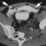
There were 42 patients with extraperitoneal bladder injuries. All were due to blunt trauma, and 92% were associated with pelvic fractures. Most were found with CT cystogram. Two patients had operative repair, probably due to the need to fix the pubic bones with hardware. 37 of the 42 were available for followup, and 22% of repeat cystograms were positive (average study done on day 9). In the studies that showed a leak, repeat cystograms were done, and they took an average of 47 days to fully heal.

Bottom line: Patients with extraperitoneal or complex intraperitoneal bladder injuries (trigone) really do need a followup cystogram before removing the bladder catheter. Those who underwent a simple repair of their intraperitoneal injury do not.

Related posts:

Reference: Cystogram follow-up in the management of traumatic bladder disruption. J Trauma 60(1):23-28, 2006.

Extraperitoneal Bladder Rupture

This injury is likely to occur in patients who have a full bladder and sustain anterior pelvic trauma that typically leads to fractures. They generally present with gross hematuria upon placement of the bladder catheter. This should prompt an abdominal CT scan with cystogram technique.

CT cystogram involves pressurizing the bladder with contrast prior to the study. This differs from the usual method of clamping the catheter and allowing the bladder to passively fill. The literature here is clear: failure to use cysto technique will miss 50% of these injuries.

The majority of extraperitoneal bladder injuries can be treated nonoperatively, and probably do not need Urology involvement. The bladder catheter is left in place 10-14 days (we do 10 days), and a repeat cystogram is obtained. If there is no leak, the catheter can be removed. If there is still some leakage, Urology consultation should then be obtained. 

There are a few cases where operative management is required:

  • There is some intraperitoneal component of bladder injury
  • Fixation of the pubic rami is required (bathing the orthopedic hardware with urine is frowned upon)
  • Failure of conservative management

Arrows in the photo show extraperitoneal extravasation of cystogram contrast.

Related posts:

Evaluation of Hematuria in Blunt Trauma

Hematuria ranges from microscopic to gross. Microscopic means blood that can only be seen with a microscope, and gross means visible to the naked eye. In trauma, we only care about gross hematuria, which ranges from the faintest of pink to the deepest red.

In trauma, gross hematuria is a result of an injury to kidney, ureter or bladder. Blunt injury to the ureter is so rare it’s reportable, so you can pretty much forget that one unless the mechanism is extreme. So you really just need to focus on kidney and bladder.

Any victim of blunt trauma that presents with visible hematuria needs to be evaluated by CT of the abdomen and pelvis with an added CT cystogram. Standard CT technique is done without a urinary catheter, or with the catheter clamped. Only 50% of bladder injuries show up with this technique.

CT cystogram is an add-on to the standard CT, and consists of the administration of contrast into the bladder which is then kept under pressure while the scan is done. Delayed slices through the pelvis after the bladder is depressurized and emptied is routine. Nearly 100% of bladder injuries are detected using this technique.

If the CT shows a renal laceration or hematoma, the patient should be admitted and managed according to your solid organ injury protocol. Kidney injuries fare better that livers and spleens, and only rarely require surgery. If no kidney or bladder injury is seen, the default diagnosis of a renal contusion is the culprit. No treatment is needed, and the patient can be discharged if no other injuries are present. The blood will clear over a few days, but may disappear and reappear a few times in the process. The patient can followup with their primary care physician in a week or two.