Category Archives: General

Guidelines for Consultants to the Trauma Service

Trauma surgeons often rely on consultants to assist in the care of their patients. Orthopedic surgeons and neurosurgeons are some of the more frequent consultants, but a variety of other surgical and medical specialists may be needed. I have found that providing a set of guidelines to consultants helps to ensure quality care and provide good communication between caregivers and patients / families.

We have disseminated a set of guidelines to our colleagues, and I wanted to touch on some of the main points. You can download the full document using the link at the bottom of this post.

In order to deliver the highest quality and most cost-effective care, we request that services we consult do the following:

  • Please introduce yourself to our patient and their family, and explain why you are seeing them.
  • Although you may discuss your findings with the patient, please discuss all recommendations with a member of the trauma service first. This avoids patient confusion if the trauma team chooses not to implement any recommendations due to other patient factors you may not be aware of.
  • Document your consultation results in writing (paper or EMR) in a timely manner.
  • If additional tests, imaging or medications are recommended, discuss with the trauma service first. We will write the orders or clear you to do so if appropriate, and will discuss the plan with the patient.
  • We round at specific times every day and welcome your attendance and input.
  • Please communicate any post-discharge instructions to us or enter in the medical record so we can expedite the discharge process and ensure all followup visits are scheduled.

Bottom line: A uniform “code of behavior” is important! Ensuring good patient communication is paramount. They need to hear the same plans from all of their caregivers or else they will lose faith in us. One of the most important lessons I have learned over the years is that you do not need to implement every recommendation that a consultant makes. They may not be aware of the most current trauma literature, and they will not be familiar with how their recommendations may impact other injuries.

Click here to download the full copy of the Regions Hospital Trauma Services consultant guidelines.

Print Friendly, PDF & Email

Serial Hemoglobin To Monitor Chest Tube Output? Huh?

I’ve already written about the (f)utility of serially monitor hemoglobin (Hgb) or hematocrit (Hct) levels when managing solid organ injury nonoperatively. What about if you are concerned with bloody output from a chest tube drainage system? Could it be of any use there?

Seems like a reasonable idea, right? Wrong. As always, think it through and do the math! Here are the questions you need to ask yourself:

  • What is the Hgb or Hct of the fluid coming out of the chest tube? At worst, it will be the same as the patient, assuming that pure, whole blood is coming out. But this is seldom the case. The fluid is usually described as “serosanguinous”, which is not very exact, but tells you that it is thinner than blood. And if it looks more like Kool-Aid, the concentration is very low indeed.
  • What is the volume in the container? Most collection systems will collect a maximum of 1 to 1.5 L of juice.
  • How fast is it coming out? These things almost never fill right in front of your eyes. It’s a slow process, with less than a few hundred ccs per shift.

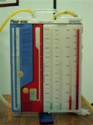
Here’s a few hundred ccs of thin drainage in a collection system. Probably decrease in Hgb value – < 0.1, which is far less than the range of lab error.

Bottom line: So now do the math. Let’s say the fluid has half the hematocrit / hemoglobin of whole blood. Losing one unit (500cc) of whole blood will generally drop your Hgb by about 1 gm, or your Hct by about 3%. If the blood is half-strength like I am proposing (and the usual drainage is typically much thinner), it will take twice as much (one liter) loss to drop the lab values by that much. This will probably come close to filling up the average collection system. If it takes a day or two or more to fill up, you are not going to see much change in their lab values. And most of the time, the blood in the system is thin like Kool-Aid, so your patient is really losing very little actual blood.

So measuring serial hemoglobin / hematocrit as you watch a hemothorax drain doesn’t make sense. Unless the output is pure blood and the system is filling up in front of your eyes, of course. In that case, a trip to the OR to fix the problem might be a better idea than doing a blood draw and sitting around waiting for the result to come back.

Related post:

Print Friendly, PDF & Email

Blood Transfusion With Component Therapy vs Whole Blood

About 40 years ago, blood banks started moving away from keeping whole blood and began separating it into components (packed cells, platelets, plasma, etc.) for more targeted use. For most uses, this is just fine. But what about trauma?

Trauma patients bleed whole blood. Doesn’t it make sense to give whole blood back? Much of our experience with massive transfusion is derived from our colleagues in the military. Two decades ago, the norm was to give 4 units of packed red cells or so, then give two units of plasma, and every once in a while slip in a bag of platelets. Our military experience seems to indicate that this 4:2:1 ratio is not optimal, and that something like 1:1:1 is better.

If you think about it, whole blood is already 1:1:1. Splitting it into components and then giving each one of them back separately seems to be a lot of extra work (and expense) to accomplish the same thing as just giving a unit of whole blood. And if you look at the purple table above, rebuilding a unit of whole blood from components isn’t nearly as good as whole blood. Plus it triples the exposure to infectious agents and antigens, since the components will usually come from (at least) three separate donors. Note that the data in the table above is true for fresh whole blood (not practical in civilian life); banked whole blood will still lose some coagulation activity. 

Is it time to think about supplying whole blood to trauma centers? And actually looking at whether the outcomes are better or not?

Print Friendly, PDF & Email

Advanced Needle Thoracostomy

In the past, I’ve written about the merits of needle vs finger thoracostomy. One of the arguments against needle thoracostomy is that it may not reach into the chest cavity in obese patients. As I mentioned yesterday, use the right needle!

Obviously, the one on top isn’t going to get you very far. The bottom one (10 gauge 3 inch) should get into most pleural spaces.

But what if you don’t have the right needle? Or what if the patient is massively obese and the longer needle won’t even reach? Pushing harder may seem logical, but it doesn’t work. You might be able to get the needle to reach to the pleural space, but the catheter won’t stay in it.

Here’s the trick. First, make the angiocatheter longer by hooking it up to a small (5 or 10cc) syringe. Now prep the chest over your location of choice (2nd intercostal space, mid-clavicular line or 5th intercostal space, anterior axillary line) and make a skin incision slightly larger than the diameter of the syringe. Now place the syringe and attached needle into the chest via your incision. It is guaranteed to reach the pleura, because you can now get the hub of the catheter down to the level of the ribs. Just don’t forget to pull out the catheter once you’ve placed the chest tube!

Related posts:

Print Friendly, PDF & Email

The Right Way to Treat Tension Pneumothorax

Tension pneumothorax is an uncommon but potentially lethal manifestation of chest injury. An injury to the lung occurs that creates a one-way valve effect, allowing a small amount of air to escape with every breath. Eventually the volume becomes so large as to cause the lung and mediastinum to push toward the other side, with profound hypotension and cardiovascular collapse.

The classic clinical findings are:

  • Hypotension
  • Decreased or absent breath sounds on the affected side
  • Hyperresonance to percussion
  • Shift of the trachea away from the affected side
  • Distended neck veins

You should never diagnose a tension pneumothorax with a chest xray or CT scan, because the diagnosis is a clinical one and the patient may die while these procedures are carried out. Having said that, here’s one:

image

The arrow points to the completely collapsed lung. Note the trachea bowing to the right.

As soon as the diagnosis is made, the right thing to do is to “needle the chest.” A large bore angiocath should be placed in the second intercostal space, mid-clavicular line, sliding right over the top of the third rib. The needle should then be removed, leaving the catheter.

The traditional large bore needle is 14 gauge, but they tend to be short and flimsy. They may not penetrate the pleura in an obese patient, and will probably kink off rapidly. Order the largest, longest angiocath possible and stock them in your trauma resuscitation rooms.

image

The top catheter in this photo is a 14 gauge 1.25 inch model. The bottom (preferred at Regions) is a 10 gauge 3 inch unit. Big difference! 

The final tip to treating a tension pneumothorax is that a chest tube must be placed immediately after inserting the needle. If the patient is on a ventilator, the positive pressure will slowly expand the lung. But if they are breathing spontaneously, the needle will change the tension pneumothorax into a simple open pneumothorax. Patients with other cardiovascular problems will not tolerate this for long and may need to be intubated if you dawdle.

Related post:

Print Friendly, PDF & Email