Category Archives: General

Trauma Centers: Optimal Resources vs Optimal Care

Since its inception, the trauma verification program for the American College of Surgeons has focused on standardizing the resources required by a hospital in order for it to become a trauma center. The book that contains the requirements for all levels of trauma center is even titled the “Optimal Resource Document.”

But is achievement of optimal resources enough? Numerous studies have shown that by adhering to these requirements and becoming a trauma center, trauma patient outcomes are improved and mortality is decreased. However, there is still considerable outcome variability across the same level of trauma centers to suggest that it might not be.

Trauma centers accomplish two things: providing a mostly standard set of at-the-ready resources, and providing care using these resources via a set of processes. Some of these processes have been codified by various organizations as practice guidelines. Are variations in the way these processes are implemented the reason for the observed variability in outcomes across trauma centers of the same level?

A paper by Shahid Shafi looked at compliance with standard trauma processes of care (T-POC) and the impact on outcome (mortality) in a single Level I trauma center in the US during a 3 year period. A total of 994 patients were studied. The authors focused on 25 process of care algorithms in 4 specific groups of common injury problems, including TBI, shock, pelvic fracture, and long bone extremity fracture.

The following interesting findings were noted:

  • 77% of the patients were eligible for at least one or more T-POC
  • Compliance with specific T-POCs varied from 10% to 99%
  • The best compliance rates (>90%) were achieved in only 3 T-POCs (blood transfusion for hypotension, intubation for low GCS, and laparotomy for GSW to the abdomen)
  • As judged by whether application of the various T-POCs actually occurred, half of the patients received only 60% of the care they needed
  • Increasing compliance with T-POCs by 10% decreased risk-adjusted mortality by 14%

Bottom line: Adoption of and adherence to standard process of care algorithms for various common clinical problems is the next step in the evolution of trauma care. Yes, a standard set of at-the-ready resources will still be required. But the verification and designation process for trauma centers will need to evolve to place more emphasis on not just having a collection of evidence based T-POCs, but on how uniformly and consistently they are properly applied.

Reference: Moving from “optimal resources” to “optimal care” at trauma centers. J Trauma 72(4):870-877, 2012.

Pop Quiz – Final Answer!

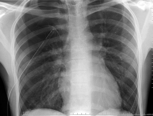
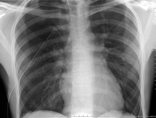
So what was wrong with that post-chest tube x-ray taken after placement for treatment of a pneumothorax?

The answer? The chest tube is not in the chest!

But wait, you say. That x-ray looks totally normal. The placement couldn’t be more perfect!

Remember, an x-ray image is a 2D representation of a 3D object. You can easily see the location of the tube in the x and y axes, and it looks great. But the z (depth) axis? You have no clue. And in this case, the tube is along the posterior chest, under the scapula. That’s a potential reason for the persistent pneumothorax.

Some readers commented that the tube could be in the lung parenchyma or a fissure. Fissure placement does occur occasionally, and may hamper the function of the tube somewhat, but it will still work. My radiology colleagues occasionally call me to warn that the tube goes through the lung parenchyma. Fortunately for the patients, this is almost never true. Just an illusion seen on the CT. It is very difficult to place a tube through normal lung. The resistance is substantial enough to make any reasonable person stop pushing. Hopefully.

Bottom line: Remember that a plain x-ray is only two dimensional. Your brain will place the objects seen on it wherever you desire. But you do not know how deep or superficial any object is without additional information. Even CT scans simulate 3D by stacking a bunch of this slices or shaded images to fool your brain. When placing a chest tube, verify its insertion point with your finger; you can follow the tube down to the chest wall and feel it vanish between the ribs. Don’t just assume you know where it’s going.

Hat tips to @CookCountyTraum, HollyT and Josh for getting it correct!

Pop Quiz! Hint Hint!

Yesterday I hit you with a chest x-ray after chest tube insertion in a young man who presented with a pneumothorax. The lung was not yet expanded (chest x-ray taken less than 5 minutes after the procedure).

So what’s wrong? I had a lot of good guesses yesterday (@ResusReview, @uclamutt, and others in the comments to name a few), but nobody quite got it. Yes, the lung is not up yet. No, the tube is not in a fissure. The person inserting the tube worked up a sweat doing it, taking about 10 minutes to get it in. But some air came out initially, and the tube rotated freely on its axis.

Any ideas? Tweet or comment! Answer tomorrow.

Pop Quiz! What’s Wrong In This Picture?

Here’s another one to challenge your skills! A young male presents to your ED after minor chest trauma with pleuritic chest pain and slight shortness of breath. A chest x-ray is obtained which shows a large pneumothorax on the right. You insert a chest tube, and the procedure goes well.

Another x-ray taken immediately after insertion looks like this:

The lung is not yet fully expanded, but the patient already feels better. 

What is wrong in this picture? Tweet or leave comments. Hints tomorrow if I don’t receive the correct answer.

But The Radiologist Made Me Do It!

The radiologist made me order that (unnecessary) test! I’ve heard this excuse many, many times. Do these phrases look familiar?

  1. … recommend clinical correlation
  2. … correlation with CT may be of value
  3. … recommend delayed CT imaging through the area
  4. … may represent thymus vs thoracic aortic injury (in a 2 year old who fell down stairs)
Some trauma professionals will read the radiology report and then immediately order more xrays. Others will critically look at the report, the patient’s clinical status and mechanism of injury, and then decide they are not necessary. I am firmly in the latter camp.
But why do some just follow the rad’s suggestions? I believe there are two major camps:
  • Those that are afraid of being sued if they don’t do everything suggested, because they’ve done everything and shouldn’t miss the diagnosis
  • Those that don’t completely understand what is known about trauma mechanisms and injury and think the radiologist does
Bottom line: The radiologist is your consultant. While they are good at reading images, they do not know the nuances of trauma. Plus, they didn’t get to see the patient so they don’t have the full context for their read. First, talk to the rad so they know what happened to the patient and what you are looking for. Then critically look at their read. If the mechanism doesn’t support the diagnosis, or they are requesting unusual or unneeded studies, don’t get them! Just document your rationale clearly in the record. This provides best patient care, and minimizes the potential complications (and radiation exposure) from unnecessary tests.
Related post: 

Reference: Pitfalls of the vague radiology report. AJR 174(6):1511-1518, 2000.