Category Archives: General

Weird DPL Case: The Answer

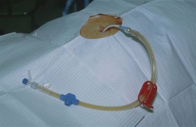
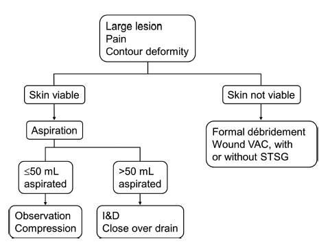
The patient underwent DPL and had an abnormal result, with weird sediment in the tubing. 

Since this is, by definition, a positive result, they were taken to the OR. The DPL catheter was left in place to help localize what was going on. This is particularly helpful if an iatrogenic injury to a hollow organ is suspected. Otherwise it may be very difficult to find a tiny puncture wound.

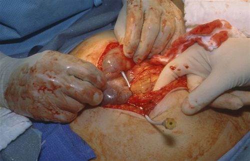
It turns out that the catheter made a beeline for the cecum, resulting in a DCL (diagnostic colonic lavage), not a DPL. The particulate material was stool!

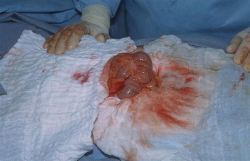
Since the catheter had been left in place, there was no contamination. It was removed under direct vision and a single stitch was placed to close the defect. Finally, a formal exploration was carried out to find the source of the patient’s abdominal pain. A low grade liver laceration was found that did not need any specific therapy. The ultimate source of her initial hypotension? Multiple long bone fractures with attendant bleeding into soft tissue. The abdomen was closed and the patient did well after fixation of her fractures.

It pays to know a little bit about DPL, even though it is seldom used these days. It can be useful, particularly when trying to rule the abdomen in or out as a source of bleeding where FAST is unavailable, indeterminate, or the result is suspect.

Check out this post for some tips and tricks on DPL:

Print Friendly, PDF & Email

Interesting Case: Part Deux

This is a continuation of yesterday’s interesting case involving an unusual DPL result. As you recall, the tap was negative, but the lavage effluent slowly began to show some particulate material.

By definition, this is a positive result, which then requires a trip to the operating room. The catheter was capped and left in place. The patient was then taken to surgery, prepped and draped. Here’s what was found:

What’s your diagnosis now? And what needs to be done about it?

Final answers tomorrow!

Print Friendly, PDF & Email

Interesting Case

Here’s an interesting trauma case, which comes from days of DPL. Although we don’t use this valuable technique very often, this one teaches an interesting lesson.

A middle aged female was involved in a high speed car crash. She was brought to the resuscitation bay as a trauma activation because the medics reported she had bilateral femur fractures, and her systolic pressures were in the 90’s. 

As you proceed through the ATLS protocol, you call for blood to supplement your resuscitation fluids, and you also find that her abdomen is tender, with some right upper quadrant guarding. The femurs are placed in traction splints. FAST is generally negative, but the right upper quadrant is equivocal.

At this point, her pressure drops again. You re-evaluate your ABCs and find nothing new. The femurs appear to be nicely reduced, and the thighs are not larger than they were when she arrived. Your surgeon is concerned that the abdomen may be the source despite the (mostly) negative FAST. Due to BP concerns, she proceeds to do a DPL.

The procedure proceeds smoothly while resuscitation with blood products takes place. There is no gross blood on the tap. A liter of saline is infused and is now freely emptying into a bag. For the first 400cc, the effluent is crystal clear. But now you start to see something.

Hmm, is it or isn’t it? Let’s take a closer look.

Yeah, that’s weird. Just of hint of some kind of tiny darkish particles settling to the bottom of the tubing. Hmmm!

So what’s happening here? And what should you do? More information tomorrow. Please comment or tweet your guesses!

Print Friendly, PDF & Email

Newest Cool Device: XStat Dressing

This new investigational device has made quite a splash during the past week. Manufactured by an Oregon company, it is designed to control bleeding, and is for use by combat medics and first responders.

Inspired by the old Fix-A-Flat expanding foam tire patch system, the XStat looks like a big syringe, and is filled with small 1cm sponges that expand rapidly when they get wet. It’s designed to stop hemorrhage in small wounds and wound tracts. Just pull back the plunger (which comes fully inserted to save space), push the unit into the wound, then hold the plunger while pulling the syringe out. This serves to leave the load of sponges in the tract and achieve rapid hemostasis.

image

It would seem that leaving a lot of tiny sponges in a wound could cause problems, especially if they are not removed at the time of definitive surgical management. However, each one is tagged with a radiopaque marker so they can be identified with xray or fluoro.

Preclinical trials have claimed to be successful, and an application has been submitted to the FDA for human use. This has the potential to save lives when bleeding gunshot wounds are encountered, especially in combat situations.

I have no financial interest in RevMedx, the manufacturer of this device.

More info: 

Print Friendly, PDF & Email

What Is: A Morel-Lavallee Lesion?

Anyone who takes care of blunt trauma has seen the Morel-Lavallee lesion (M-L). Here’s an obvious one because it’s acute:

The M-L lesion is essentially a closed degloving injury in which the skin remains intact. The subcutaneous tissue is sheared off of the underlying fascia, and typically blood accumulates in the potential space that is created. This picture shows a less acute lesion; the bruising and ecchymosis on the surface have resolved. Note the collection on the lateral thigh:

These injuries may take a very long time to resolve and may leave some residual deformity. The definitive management has never been very clear: needle drainage vs incision, timing, compression wraps, etc.

The Mayo Clinic reviewed their 8 year experience with 87 of these lesions to try to shed some light on proper management. They treated their patients in four different ways: needle drainage, incision and drainage, compression wraps, and debridement with vacuum drainage devices. Here are the factoids from their study:

  • Motor vehicle crash was the most common etiology for this lesion, which makes sense due to the energy needed to shear the tissues
  • The most common locations were thigh, hip and flank
  • The incidence of pre-existing conditions that might influence outcome (diabetes, obesity, smoking history, use of anticoagulants) did not seem to influence outcomes
  • Lesion location did not change the recurrence rate (even over joints)
  • Aspiration suffered the highest recurrence rate (56%) vs only 15-19% in the other groups
  • Aspiration of more than 50cc of fluid was more common in lesions that recurred (83%) vs those that did not (33%)

Their experience led them to develop the following practice guideline:

Bottom line: The Morel-Lavallee lesion can be challenging to treat. Although this study has limited numbers, it provides enough guidance to suggest a consistent way of managing it. I recommend adopting this algorithm to provide a standard pathway for dealing with it.

Reference: The Mayo Clinic experience with Morel-Lavallee lesions: establishment of a practice management guideline. J Trauma 76(2):493-497, 2014.

Print Friendly, PDF & Email