All posts by The Trauma Pro

Submental Intubation – The Video!

Yesterday, I described a novel technique for providing a secure yet short-term airway tailored to patients who can’t have a tube in their mouth or nose. Patients undergoing multiple facial fracture repair are probably the best candidates for this procedure.

A picture may be worth a thousand words, but a video is even better. Please note that it is explicit and shows the blow by blow surgical procedure. Of note, it is a quick and relatively simple advanced airway technique. Note the cool music!

YouTube player

Related post:

By Request: Submental Intubation

I keep getting requests regarding this technique, so I’m reposting  this updated article today and tomorrow.

Here’s one of the weirder procedures I’ve seen in some time. Imagine that you need a definitive airway, but you can’t use the face for some reason (mouth or nose). The usual choice would be a tracheostomy, right? But what if you only need it for a few days? Typically, once placed, trachs must be kept for a few weeks before decannulation is safe.

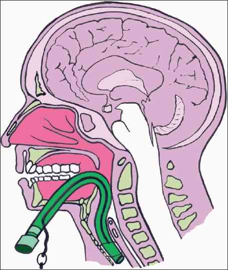
Enter submental intubation. This technique involves passing an endotracheal tube through the anterior floor of the mouth, and then down the airway. This leaves the facial bones, mandible, and skull base untouched.

tumblr_inline_mvds1c14ye1qa4rug1


The technique is straightforward:

  • After initially intubating the patient  orotracheally, a 1.5cm incision is created just off the midline in the submental area under the chin.
  • Using a hemostat, all layers are penetrated, entering the oropharynx just lateral to the tongue.
  • A 1.5cm incision is then made at the puncture site, parallel to the gum line of the lower teeth.
  • The ET tube is removed from the ventilator circuit, and the connector at the proximal end of the tube is removed.
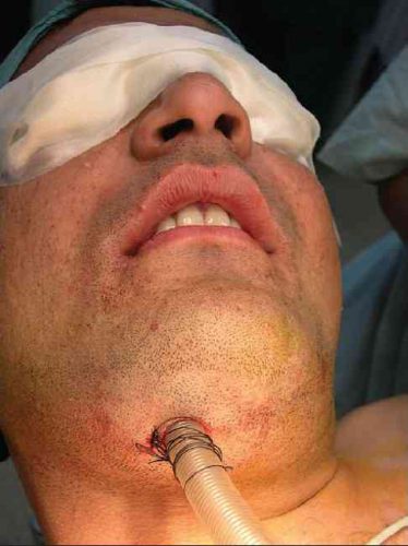
  • The hemostat is placed through the chin incision again. The proximal end of the ET tube is curled into the oropharynx and grasped with the hemostat, then pulled out through the skin under the chin, leaving the distal (balloon) end in the trachea.
  • The connector is reinserted, and the tube is then hooked up to the anesthesia circuit again.
  • The tube is then secured using a stitch under the chin.

After a final position check, the surgical procedure can commence. Cool!


There are a number of variations on this technique, so you may encounter slightly different descriptions. The tube can be pulled at the end of the procedure, or left for a few days to ensure safe extubation, if needed.

tumblr_inline_mvdsm0zd8b1qa4rug1

A small series of 10 patients undergoing this technique was reviewed, and there were no short or long term problems. Scarring under the chin was acceptable, and was probably less noticeable than a trach scar.

Bottom line: This is a unique and creative method for intubating patients with very short-term airway needs while their facial fractures are being fixed. Brilliant idea!

Tomorrow: Submental intubation – the video!

Reference: Submental intubation in patients with panfacial fractures: a prospective study. Indian J Anaesth 55(3):299-304, 2011.

Photo source: internet

Update: The Rectal Exam In Trauma Continues to “Pass”?

This topic continues to come up from time to time. I still see trauma programs that perform the good, old-fashioned digital rectal exam on nearly every trauma patient. But is it really necessary?

In the not so distant past, it was standard operating procedure to perform a digital rectal exam in all major trauma patients. The belief had always been that valuable information about blood in the GI tract, the status of the urethra, and the neuro exam (rectal tone) could be gleaned from this exam.

Unfortunately, a finger in the bum also serves to antagonize or even further traumatize some patients, especially those who may be intoxicated to some degree. On a number of occasions I have seen calm patients become so agitated by the rectal exam that they required intubation for control.

So is it really necessary? A study in 2001 conducted over a 6 month period (1) showed that the rectal exam influenced management in only 1.2% of cases. The authors felt that there was some utility in 3 special cases:

  • Spinal cord injury – looking for sacral sparing
  • Pelvic fracture – looking for bone shards protruding into the rectum
  • Penetrating abdominal trauma – looking for gross blood

A more recent 2005 study (2) was also critical of the rectal exam and found that using “other clinical indicators” (physical exam and other diagnostic study information) was at least equivalent, changing management only 4% of the time. They concurred with the first two indications above as well.

And what is the best patient position for the exam? I continue to see people try to do it when the patient is in the logroll position! This is substandard for two reasons:

  • It is not a stable position, and no one likes a finger in their butt. Awake patients will squirm and withdraw, defeating any attempt at spinal precautions.
  • It’s not ideal for the examiner, either.  Access to the male prostate is subpar because the examiner’s finger is generally pointed posteriorly, away from this organ. In order to rotate anteriorly, the examiner must spin around, putting “reverse English” (billiards reference) on their arm.

To do a proper rectal exam when indicated, make sure the patient is supine, warn them that you are going to do it, and use the same hand as the side of the patient you are standing on. Right side, right hand to avoid the “reverse English” thing again.

Bottom line: For most major trauma patients, the rectal exam is not worth the patient aggravation it causes. I still recommend it for the 3 special cases listed above, however, as there are no equivalent and effective exams for these potentially serious patient problems. And remember, DON’T do it while the patient is in the logroll position

References:
1. Porter, Urcic. Am Surg. 2001 May;67(5):438-41.
2. Esposito et al. J Trauma. 2005 Dec;59(6):1314-9.

The May 2019 Trauma MedEd Newsletter Has Been Released!

Here’s the most recent newsletter that was released at the end of May; the topic is Spleen Injury Update.

In this issue, I cover:

  • Update To Spleen Injury Scaling / Grading
  • Overwhelming Post-Splenectomy Infection
  • Spleen Vaccines
  • Early Mobilization In Solid Organ Injury

To download the current issue, just click here! Or copy this link into your browser: http://bit.ly/TME201905

The next newsletter will contain an analysis of the results of my Trauma PI Coordinator survey. It will be released only to subscribers at the end of June.  This one will not be released on the blog, but will be available as a subscriber sign-up bonus beginning next month.  Click this link right away to sign up now and/or download back issues.

Got a suggested theme for later issues? Just let me know what you’d like to read about by emailing or leaving a comment here.

Fat Embolism Syndrome And Orthopedic Surgery

Regardless of the exact mechanism for the development of fat embolism syndrome, in trauma it most commonly occurs when the medullary (bone marrow) cavity of a long bone is violated. This occurs first when the bone is fractured, and again when it is instrumented for fixation. The initial shower of emboli cannot be prevented. However, ongoing emboli can be reduced with early fixation. This can be in the form of a good splint, or surgical external or internal fixation.

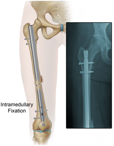
One type of internal fixation, intramedullary (IM) nailing, has been associated with embolism and FES for some time. This technique was introduced 80 years ago and has been refined significantly since. Here is a picture of a femur with an IM nail.

The nail is inserted proximally near the greater trochanter. The marrow cavity is first reamed to make insertion of the nail easier. This causes a number of changes in the physiology of and pressures within the marrow cavity. Pressure increases during the initial reaming, and hits a peak when the reamer enters the distal fragment. Once complete, there are no further increases as the nail is inserted. However, these pressure changes alter medullary blood flow and allow emboli to enter the venous system.

Reaming is actually beneficial in several ways. It simplifies and shortens the surgical procedure. And in animal models there is evidence that bone debris from the reaming process collects at the fracture site, creating an autograft that may improve healing.

A surgical group in Ireland has been using a novel technique for lavaging the marrow cavity during fixation for several years. Once the bone is entered proximally, a cut piece of suction tubing is inserted into the end of the bone. Suction is then applied for 2-3 minutes. The procedure continues, including reaming, then the suction procedure is repeated. Unfortunately, FES is uncommon, so it is difficult to judge whether their technique really works. The authors believe it is safe, but recommend formal studies to prove efficacy.

Use of an additional venting hole between the trochanters has also been studied in a small randomized trial. This allows for drainage of marrow during the reaming process, reducing any pressure rise. The number of embolic events detected using transesophageal echo was significantly reduced in the vented group (20% vs 85% of patients).

Tomorrow, prevention and treatment of fat embolism syndrome.

References:

  1. A Simple and Easy Intramedullary Lavage Method to Prevent Embolism During and After Reamed Long Bone Nailing. Cureus 9(8):e1609, Aug 2017.
  2. Relevance of the drainage along the linea aspera for the reduction of fat embolism during cemented total hip arthroplasty. A prospective, randomized clinical trial. Arch Ortho Trauma Surg 119:146, 1999