Pediatric Solid Organ Injury Management: It’s About Time!

There was an interesting article released in the Journal of Pediatric Surgery in May about spleen and liver injury management in children. It’s interesting because if you just look at the title, you might just skip over it. The title suggests that it describes reducing scheduled phlebotomy in kids who are undergoing solid organ injury management. But the real meat of this article has to do with the protocol they are using to treat the children.

Nonoperative management of these injuries in children started becoming popular 40 years ago (!). But for decades, everyone put their own spin on how to do it. Bed rest for a week (or more). NPO for days! Limited physical activity for extended periods. Then the American Pediatric Surgery Association (APSA) published a set of guidelines about 15 years ago that took some of the guesswork out of it.

Although nonoperative management of these injuries in kids preceded its adoption in adults by a nearly two decades, it has languished in the APSA format for quite some time. Many pediatric surgeons still use these guidelines, even though adult spleen and liver injury management have advanced to shorter and more streamlined care.

We adopted a solid organ management guideline at Regions Hospital over ten years ago, and have made a few minor tweaks over the years. Nowadays, our grade I-III injuries can be home as early as 36 hours after admission, and frequently are. Grades IV and V are eligible to be discharged after just 24 more hours if they have no other injuries to keep them in the hospital.There are very rare failures.

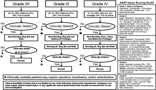
I’ll detail the factoids about the phlebotomy part of this paper in tomorrow’s post. But I do want to show you the more aggressive protocol the authors are using (one of whom authored the original APSA guideline).

Here it is:
image

Bottom line: Note how quickly children are allowed to get up, eat, and get out of the hospital using the “new” protocol. Many adult centers have been using similar ones for years. It’s nice to see that adult and pediatric protocols are finally beginning to converge. After all, we figured out our current adult management based on our experience with kids 30 years ago! 

Related posts:

Reference: Reducing scheduled phlebotomy in stable pediatric patients with liver or spleen injury. J Ped Surg 49(5):759-762, 2014.

Another Way To Treat Tension Pneumothorax

Kenji Inaba and colleagues have done a lot of work on tension pneumothorax (tPTX) in the past few years. They’ve looked for the best devices and the best positions on the chest to quickly and effectively treat this emergency. Now, they’ve published a study on using what looks like a “better mousetrap” for relieving tension physiology.

Previous work from this lab has shown that up to a quarter of needle thoracostomies fail within 5 minutes due to mechanical reasons. This leaves a small window for insertion of the real chest tube. And even though much of the pressure may be relieved, a significant amount of air may be left in the chest, impeding recovery from PEA arrest.

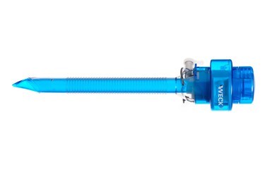
They looked at the use of a 5mm laparoscopy port for relief of tension pneumothorax in Yorkshire swine. The exact size of the pigs was not listed, but these animals weigh 25 pounds at 6 weeks of age, and the pictures in the article show a reasonable sized animal. I’m not sure they were 70kg, though.

Here are the factoids:

  • Five animals were used, and 30 episodes of tPTX and 27 episodes of PEA arrest from tPTX
  • Tension pneumothorax was created by insufflating the chest with CO2 using a 10mm laparoscopic trocar
  • tPTX was completely relieved by insertion of the 5mm trocar in 100% of trials, with all physiologic measures returning to baseline within 1 minute
  • Circulation was restored to normal within 30 seconds in 100% of trials
  • There was no damage to heart or lung from trocar placement in any of the 5 animals

image

Bottom line: Once again, Inaba and crew have added some interesting tidbits to our knowledge base. You already know I’m not a fan of animal studies like this, but this one lays the ground work for some work in humans. We still need to know how the “usual American body habitus” will affect the use of this device. The only downside is the expense of the trocar, which is a lot more than a simple long needle. But if it is as efficacious in humans as it is in pigs, it may be worth it!

Related posts:

Reference: Standard laparoscopic trocars for the treatment of tension pneumothorax: A superior alternative to needle decompression. J Trauma 77(1):170-175, 2014.

Procedural Complications: Residents vs Advanced Practice Providers

With the implementation of resident work hour restrictions 10 years ago, resident participation in clinical care has declined. In order to make up for this loss of clinical manpower and expertise, many hospitals have added advanced clinical providers (ACPs, nurse practitioners and physician assistants). These ACPs are being given more and more advanced responsibilities, in all clinical settings. This includes performing invasive procedures on critically ill patients. 

A recent study from Carolinas Medical Center in Charlotte NC compared complication rates for invasive procedures performed by ACPs vs residents in a Level I trauma center setting.

A one year retrospective study was carried out. Here are the factoids:

  • Residents were either surgery or emergency medicine PGY2s
  • ACPs and residents underwent an orientation and animal- or simulation-based training in procedures
  • All procedures were supervised by an attending physician
  • Arterial lines, central venous lines, chest tubes, percutaneous endoscopic gastrostomy, tracheostomy, and broncho-alveolar lavage performances were studied
  • Residents performed 1020 procedures and had 21 complications (2%)
  • ACPs performed 555 procedures and had 11 complications (2%)
  • ICU and hospital length of stay, and mortality rates were no different between the groups

Bottom line: Resident and ACP performance of invasive procedures is comparable. As residents become less available for these procedures, ACPs can (and will) be hired to  take their place. Although this is great news for hospitals that need manpower to assist their surgeons and emergency physicians, it should be another wakeup call for training programs and educators to show that resident education will continue to degrade.

Related posts:

Reference: Comparison of procedural complications between resident physicians and advanced clinical providers. J Trauma 77(1):143-147, 2014.

The Newest Trauma MedEd Newsletter Is Available!

After taking a travel break last month, it’s back! The latest edition of
the Trauma MedEd newsletter is now available for download. The subject is Abdomen. Included are articles on:

– How to close an abdominal stab laparoscopically
– FAST is FAST and FAST is last!
– FAST exam in children
– Performance improvement for FAST
– DPL: a dying art?
– Less morbidity from negative trauma laparotomy?

The web link to this month’s issue is http://bit.ly/TME-06-2014.

You can view and download back issues at http://www.TraumaMedEd.com

Liver Function Testing After Hepatic Injury

The liver is one of the two most commonly injured solid organs after blunt trauma. There are a variety of ways to manage solid organ injury, and many trauma centers are adopting solid organ injury protocols to streamline and improve care. I am occasionally asked whether there is a place for liver function testing after hepatic injury. 

In a previous post (see below), I cited some old literature refuting this idea. A more recent paper has now tried to answer this question. They retrospectively reviewed 3 years of data on patients admitted to a large hospital in Jiangsu, China. Only patients with blunt liver injury were included. They were interested to know if liver function testing helped identify the presence and severity of injury.

Here are the factoids:

  • 182 patients who had blunt abdominal injury and liver function testing were identified in their registry (AST, ALT, GGT, Alk PHos, LDH, bili)
  • 90 patients had liver injury and 92 did not
  • Grade of liver injury was fairly evenly distributed, with a few less grade IV and V
  • Elevated LFTs accurately predicted the presence of a liver injury. ALT > 57 U/L was the most accurate predictor.
  • There was no correlation between LFT values and severity of liver injury

Bottom line: Basically, routine liver function testing after blunt abdominal trauma is a waste of time. And obtaining LFTs after known liver injury is an even greater waste of time. You know your patient has the injury, and you know the grade from the CT scan you obtained (hopefully). And from personal experience, there is absolutely no value in “trending” liver functions to see how the liver is healing. If the patient develops an unexpected clinical finding at some point (new pain, jaundice, fever), then you may wish to order laboratory or imaging studies to help determine if a complication is developing.

Related posts:

Reference: Role of elevated liver transaminase levels in the diagnosis of blunt liver injury after blunt abdominal trauma. Experimental and Therapeutic Medicine 4(2):255-260, 2012.