Trauma Surgery Tip: How To See The Unseeable

Let me present a scenario and first see how you might solve this problem.

A young man presents with a gunshot to the abdomen in the right mid-back. He is hemodynamically stable, and you get a chest xray. It shows a small caliber slug in the right upper quadrant, but no hemo- or pneumothorax. He has peritoneal signs, so you whisk him off to the OR for a laparotomy.

As you prep the patient for the case, you can feel a small mass just above the right costal margin. You incise the area and produce a 22 caliber bullet. Of course, you follow the chain of evidence rules and pass it off for the police. As you explore the abdomen, it appears that there are no gross injuries. You are concerned, however, that there may be an injury to the diaphragm in proximity to the bullet.

So here’s the question: how can you visualize the diaphragm in this area? The bullet was located below the right nipple. But the diaphragm in this area is covered by the liver, and is parallel to the floor. You can’t seem to feel a hole with your fat finger. But short of putting your whole head in the wound, you just can’t get a good angle to see the area in question.

How would you do it? Please tweet or leave comments with your suggestions. I’ll provide the answer(s) tomorrow!

The Next Best Postop Analgesia – Ice Packs?

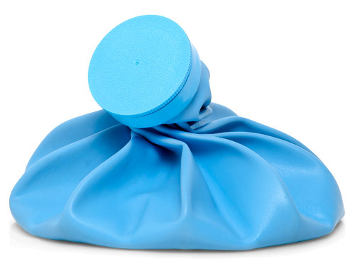
As usual, the simplest things are often the best. A recent paper looked at the newest and greatest “drug” to use for providing postoperative analgesia: the good, old-fashioned ice pack!

This concept is obviously not new. Cold is known to quiet inflammation, which is inevitable when tissues are incised. Athletes and their trainers have used ice packs forever. Surgical studies have evaluated their use in orthopedic extremity procedures as well as hernia repairs.

The current paper, from several surgical departments at Emory in Atlanta, randomly allocated patients to have an ice pack placed on their laparotomy incision. Only patients undergoing open abdominal procedures were included. Ice packs were maintained in place for 24 hours, and were then allowed as long as the patient wanted it. Pain, as judged by the analog pain scale, narcotic use, and hospital length of stay were measured. A power analysis was actually performed, and the number of patients required to detect a 15% difference were enrolled (!).

Here are the factoids:

  • 55 patients were enrolled, and were truly randomized
  • Most operations were for pancreatic, gastric, liver, and colorectal cancers
  • The usual demographics of the two groups were identical
  • Pain score was decreased as measured twice later in the day on day 1, and once on day 3
  • Narcotic use was lower on day 1
  • Length of stay was the same for both groups
  • Patients in the cryotherapy group requested to keep the ice packs for an average 2.75 days. None requested removal at the end of day 1.
  • Most stated that they would request an ice pack the next time they had surgery

Bottom line: For once, a nicely done study! Simple and to the point. It reinforces the concept that cheap and simple can still be good. The ice packs in this study were plain old refillable bags filled with ice cubes, not fancy gel or chemical packs that cost lots of money. And the decrease in narcotic use is huge! The side effects of these drugs (constipation, urinary retention, allergic reaction, etc.) create the need for interventions that introduce another whole world of complications.

Consider adding the simple old ice pack to your armamentarium of postop pain relief. But remember, you’ve got to start it as early as possible for best effect, ideally as the surgical dressing is placed.

Related posts:

Reference: Ice Packs Reduce Postoperative Midline Incision Pain and Narcotic Use: A Randomized Controlled Trial. JACS 10.1016/j.jamcollsurg.2014.03.057, 2014.

A Cool Way To Look At Injury Data

Governmental agencies everywhere collect trauma related data. The US federal government maintains a number of databases, such as the Fatal Accident Reporting System (FARS), the Census of Fatal Occupational Injuries (CFOI) and many others. States collect similar but smaller datasets. Even towns and municipalities collate injury information in the form of prehospital run sheets.

But reams of data are of no use unless you can learn something from it. Unfortunately, most of this data is tucked away in database management systems, or in some cases just stacks of paper forms locked up somewhere. In order for humans to make sense of it and do useful things with it, we need to transform it into forms that we can easily interpret and make sense of. 

Fortunately, there are lots of visual, electronic tools available to help us do just that. One of the most helpful tools is the programmable geographic information system (GIS). An example of this is Google Maps. Most of us have used this or a similar tool in some form, usually to get directions from here to there. But you may not be aware that Google provides a programming interface so a savvy user can place any type of geography-related data on the map, creating what is called a mashup.

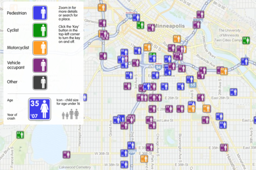
Imagine crossing the FARS database, which contains extensive data points on every fatal road accident in the US, with a mapping system. This would allow creation of a map showing where every person lost their life in a road accident, along with additional pertinent information about the event. A great example of this is demonstrated below. It was created by ITO World Ltd., based in the UK. They crossed fatality information with geographic map data in both the US and the UK.
image

This map shows fatal road events around Minneapolis from 2001 to 2009. The type of event (pedestrian struck, motor vehicle crash, etc.) is displayed along with age, year and sex. It is movable and zoomable so it can be viewed it in great detail. Click on the map above to open a new window to the full map.

Bottom line: Using trauma data / map mashups is a great way to visualize complex information. It also allows us to plan meaningful prevention activities based on local information (a requirement for ACS trauma center verification). Imagine looking over such a map of your city, and identifying a cluster of pedestrian fatalities. Then you notice that this cluster is 2 blocks away from an elementary school. This could prompt you to work with the school to implement automobile awareness programs for the children, have the city review signage and obstructions to view in the area, and optimize the number and placement of crossing guards. Then redo the map afterwards to judge the impact. Wow!

Website: http://map.itoworld.com/road-casualties-usa#fullscreen 

Reference: Using geographic information systems in injury research. J Nurs Scholarsh 39(4):306-311, 2007.
New Technology: The Bruise Suit

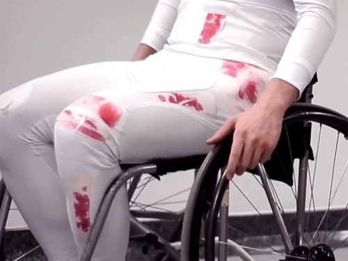
Here’s an interesting new product. It’s called the “bruise suit”, and it was designed by some students at the Imperial College of London. The purpose of the suit is to visually indicate that enough force has been applied to potentially cause injuries.

image

It was initially designed to help Paralympic athletes detect when they’ve encountered enough force to cause injuries that they are unable to feel. It uses a pressure-sensitive industrial film developed by Fuji that changes color based on the compressive force applied. It gets darker as the force increases. 

This product is currently in the concept phase, meaning that it will be some time before it hits the market. However, it’s a great idea that has implications for athletes playing contact sports and rescue professionals, to name a few. We’ll see how it develops!

Knife To The Back – The Conclusion!

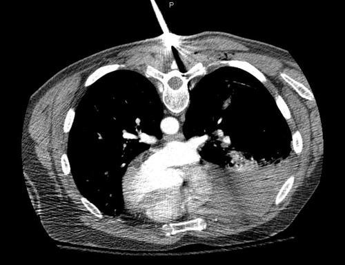
To summarize: stab to the back, prone position, stable vitals, awake and alert and breathing easily. The patient had a chest xray which showed some likely hemothorax. He was sent to CT (prone) and the image obtained looked like this:

They key points to note are:

  • The injury is completely above the diaphragm. No need to worry about an intra-abdominal problem.
  • The amount of hemothorax is moderate. It is not enough to mandate a thoracotomy. At least for now.
  • There is a significant pneumothorax. You can’t see it due to the windows used, but the lung has separated from the chest wall by about 3cm.
  • The track of the knife was directed laterally.
  • No significant vascular structures were involved, and there is no contrast extravasation.

Final management: The patient was returned to the ED, and the knife was deftly removed and processed properly as evidence. The patient was then turned supine and a 40 Fr chest tube was inserted using procedural sedation. About 400 cc of blood was drained and reinfused. A repeat chest xray was obtained, which showed some residual hemothorax and near resolution of the pneumothorax. He was then admitted for frequent vital signs and drainage measurements for two shifts. Afterwards, he was placed in our chest tube management protocol. The tube was removed and he was discharged two days later. There were no complications.