ACS vs State Trauma Verification / Designation

In the US, there are two major methods for gaining recognition as a trauma center: verification by the American College of Surgeons (ACS), and designation by a state or county authority. ACS verification is fairly rigorous and standardized, and is applied uniformly across the US. State or county designation is not nearly as uniform, with significant variability in the application of criteria, frequency of site visits, and quality of reviewers. A few states allow dual site visits which result in both ACS verification and state designation, but in general the processes are different.

A recent paper from the University of Pittsburgh sought to quantify some of these differences by using mortality as a barometer of trauma center quality and hence, verification/designation quality. It analyzed information from the 2007-2008 dataset of the National Trauma Data Bank. It specifically compared outcomes from ACS Level I and II centers to state designated centers that applied for but did not obtain ACS verification. Robust statistical models were used to address the usual shortcomings of using large datasets. Observed vs expected mortality was calculated using standard methodology. Here are the key results:

  • Over 900,000 records were analyzed (!) from a mix of academic, community, large, and small hospitals. Most (92%) were nonprofits.
  • There was no difference in survival outliers (either unexpected saves or unexpected deaths) in Level I ACS vs state trauma centers.
  • A significantly higher number of unexpected mortalities occurred in state designated Level II centers compared to ACS Level IIs. There was no difference in unexpected saves.
  • ACS verification was an independent predictor of survival at Level II centers

The equivalence in survival at Level I centers is likely due to the application of rigorous designation criteria by states for these flagship centers. Additionally, they usually attract personnel who are keenly interested in and dedicated to trauma care. Many state Level II centers have more variability in terms of commitment and resources. The surgeons may not be able to commit as much of their time to trauma care as at Level I centers. Other key resources may not be as readily available.

Bottom line: This study matches with my observations of both ACS and state trauma centers. Level I criteria are fairly rigorous for both, but there is much more variability in Level II criteria. Unfortunately, this study suggests that this situation is not good for our patients. It is not enough for states to just adopt ACS criteria into their own designation programs. Regular, standardized and rigorous reviews are important to ensuring quality.


Reference: American College of Surgeons trauma center verification versus state designation: Are Level II centers slipping through the cracks? J Trauma 75(1):44-49, 2013.

“Pull The Tube Back 2 Centimeters”

How often do trauma professionals hear that? Patients intubated in the ED (or before) almost universally have a chest x-ray taken to check endotracheal tube position. And due to variations in body habitus (and sometimes number of teeth), the tube may not end up just where we want it. So look at how deep or shallow it is and adjust it by the number of centimeters out of the correct position it should be, right?

Not so fast! A small, prospective study from Yale looked at endotracheal tube adjustment in ICU patients using tube markings and the patients incisors. Their “ideal” tube position has the tip between 2 and 4 cm from the carina. Any patients with an ET tube outside these parameters was included in the study. Here are the interesting tidbits:

  • There were only 55 patients who met criteria for the study. No denominator information was give, so we can’t tell how good or bad the intubators were initially.
  • Most tubes that needed adjustment were too far out. The median starting position was at 7cm above the carina (!),
  • A smaller number were too deep (median position 0.7cm). These were mostly in women.
  • The usual intended adjustment was 2cm. The actual distance moved after manipulation was half that (1.1cm).

Bottom line: Endotracheal tube repositioning based on tube markings at the incisors is not as accurate as you may think. Patient body habitus and reluctance to pull a tube out too far probably are factors here. So be prepared to readjust a second time unless you intentionally add an extra centimeter to your intended tube movement.

Related post:

Reference: Repositioning endotracheal tubes in the intensive care unit: Depth changes poorly correlate with postrepositioning radiographic location. J Trauma 75(1):146-149, 2013.

Pop Quiz: Final Answer

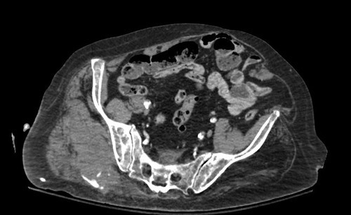
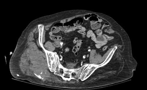
The quiz presented a single slice from a pelvic CT, asking for mechanism, age and diagnosis.

So here’s how to do it. First, what’s wrong in the picture? A number of you picked this one out. The right buttock area has a large hematoma present, and there is a contrast blush posteriorly. This is an injury to the gluteal artery with active extravasation.

Next, look at the rest of the image. Not a whole lot to see, but there is significant calcification of pelvic arteries. So this patient is elderly.

Finally, what happened? Elderly people who have gluteal artery injuries are typically pedestrians struck by a car. Most other blunt mechanisms don’t have enough energy to do this.

And what about treatment? Sometimes in younger patients, the bleeding will stop, but don’t count on it. And in the elderly, it almost never does. Angioembolization is the best way to treat this problem, because surgical exploration is generally a bloody mess.

Piece of cake! Congrats to the readers who got this one right!

I’m writing a novel and one of my characters gets stabbed in the lower region of the abdomen. I don’t want him to die or be incapacitated for a long time, so the wound can’t be too deep. The novel is set in a post-apocalyptic world so going to a hospital is not an option. What I want to know is: 1) How deep can the knife go without hitting anything vital?; 2) What do his friends need to do to help him (stop the bleeding, patch the wound…)?; 3) Would he be able to move/walk? Thanks in advance.

Great questions, and I’d love to answer in detail. Please contact me by email (see my profile at the side of the page) so we can chat!

Pop Quiz! Hints

So far, no correct answers to all three questions from yesterday’s pop quiz! Given the scan below, I want to see if you can figure out:

  • Mechanism of injury
  • Age range
  • Diagnosis

And bonus points for the correct answer on how to treat the problem!

Hint: You can tell the age by the appearance of the blood vessels in the pelvis. And the bright areas outside the pelvis may not be calcium. This should give you enough information to figure out the rest.

Tweet or comment your results!