Tag Archives: retained object

What Does A Retained Surgical Sponge Look Like?

Surgeons and surgical residents rarely see these. And because it’s so uncommon, they frequently don’t recognize the telltale findings on radiographic studies. The TSA runs into the same problem in screening passengers for weapons and other hazards at airports. But it’s the bane of any surgeon’s existence. And it’s a major reason why OR personnel take such great pains to account for everything in the room. It is a catastrophe, and always a preventable one, when some piece of equipment goes missing and ends up left inside a patient.

A number of methods have been developed to try to eliminate this problem. They include careful counts, having someone record anytime anything is placed inside, x-rays, and most recently, RFID tags.

After counting, x-ray is the most common way to try to find missing objects. One would think that these foreign bodies would be easy to see. Metallic instruments are rather easy to spot. But many trauma professionals, even those who work in the OR, have never seen what a positive image of a sponge actually looks like. So here they are. You should never miss one on an xray now.

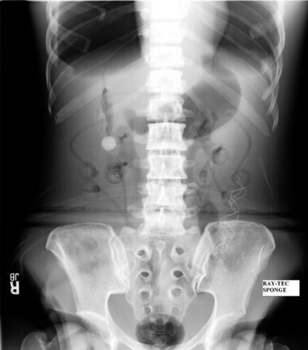
Surgeons typically use two types of sponges in the OR: Ray-Tec sponges and standard lap pads. Ray-Tecs look like a 4×8 piece of gauze with a mysterious blue string woven throughout it. The string is the only part that shows up on x-ray, and it is very thin and somewhat hard to see. Here are some Ray-Tec sponges outside the body:

And here’s one that was left inside. Note the little squiggle in the left lower quadrant and how easy it is to overlook.
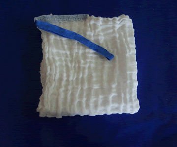
On the other hand, a laparotomy pad is a 4×4 folded cloth pad that unfolds into a larger pad. It has a blue radiopaque tag sewn in the corner, extending along one edge of the pad. Here’s what they look like outside the body:
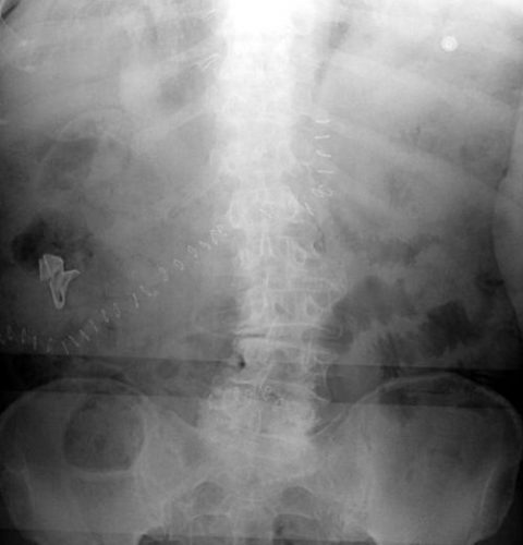
And here’s one inside a patient. Note the irregular object in the right upper quadrant. Many times the tag is scrunched up and doesn’t look like one.
Bottom line: It’s important for anyone who works in the OR on any body part to be familiar with the appearance of these tags on x-rays. Since it’s generally impossible to get accurate counts before or after a trauma procedure, always image the involved body cavity looking for these telltale signs before closing the patient.

Note: These images  were taken from the internet. Patients were not treated at Regions Hospital.
The Final X-Ray In Damage Control Surgery

Damage control surgery for trauma is over 20 years old, yet we continue to find ways to refine it and make it better. Many lives have been saved over the years, but we’ve also discovered new questions. How soon should the patient go back for definitive closure? What is the optimal closure technique? What if it still won’t close?

One other troublesome issue surfaced as well. We discovered that it is entirely possible to leave things behind. Retained foreign bodies are the bane of any surgeon, and many, many systems are in place to avoid them. However, many of these processes are not possible in emergent trauma surgery. Preop instrument counts cannot be done. Handfuls of uncounted sponges may be packed into the wound.

I was only able to find one paper describing how often things are left behind in damage control surgery (see reference below), and it was uncommon in this single center study (3 cases out of about 2500 patients). However, it can be catastrophic, causing sepsis, physical damage to adjacent organs, and the risk of performing an additional operation in a sick trauma patient.

So what can we do to reduce the risk, hopefully to zero? Here are my  recommendations:

  • For busy centers that do frequent laparotomy or thoracotomy for trauma and have packs open and ready, pre-count all instruments and document it
  • Pre-count a set number of laparotomy pads into the packs
  • Use only items that are radiopaque or have a marker embedded in them. This includes surgical towels, too!
  • Implement a damage control closure x-ray policy. When the patient returns to OR and the surgeons are ready to begin the final closure, obtain an x-ray of the entire area that was operated upon. This must be performed and read before the closure is complete so that any identified retained objects can be removed.

Tomorrow, a sample damage control closure x-ray.

Related post:

Reference: Retained foreign bodies after emergent trauma surgery: incidence after 2526 cavitary explorations. Am Surg 73(10):1031-1034, 2007.

Retained Foreign Objects After Penetrating Injury

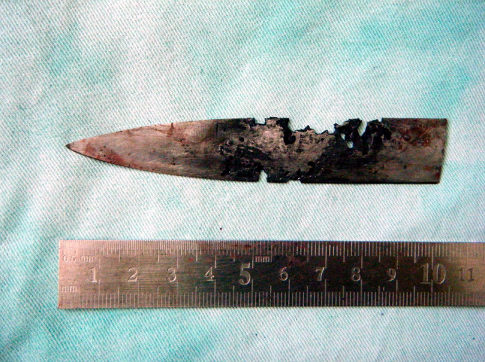
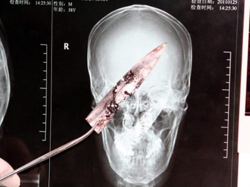
A Chinese man was in the news a few years back after having a four inch knife blade removed from his head. It had been there for four years!  The knife blade broke off after he had been stabbed under the chin. Unfortunately, he was unaware that any part of the knife had been retained. It remained partly within the nasopharynx and the tip came to rest behind his left eye. His symptoms included headaches, stuffy nose and bad breath. The picture below shows the badly corroded blade in front of some of his radiographic images.

See the video at the bottom of this post for more details and images.

knife-in-head

What is the best way to deal with a problem like this? Here are some practical tips:

First, get in the habit of imaging any body part with a penetrating injury. Retained objects can be as simple as gravel or as complicated as the knife blade above. And remember, some patients who have been stabbed present with a simple laceration but don’t want to tell you how they got it. Image before you close it!

Next, don’t remove it. This is common knowledge, but innocent looking objects (pencils, nails) can penetrate arteries and keep them from bleeding while embedded. Unpleasant and sometimes fatal bleeding can ensue if pulled out.

If you do not have specialists versed in the body regions involved in the injury, transfer immediately with the object secured in place. For objects penetrating minimally complex areas like the extremities, surgeons may opt to carefully remove it in the emergency department, or may elect to do so in the operating room.

Injuries to complex areas should undergo high resolution CT scanning so that 3D reconstruction can be performed if needed. The surgical specialists can then plan the operative approach. This is dictated by the anatomy of the area(s) involved and the architecture of the object (think about hooks and barbs). For objects located near critical areas, an operative exposure must be selected that provides access to all portions of it, and allows for rapid vascular control if needed.

YouTube player