Tag Archives: compartment syndrome

Gluteal Compartment Syndrome

Compartment syndromes can occur virtually anywhere a muscle group is surrounded by relatively unforgiving soft tissue. In trauma, these classically involve the calf, forearm, and occasionally the thigh compartments. But they are occur unsuspected in the less common areas they can easily be missed, leading to significant morbidity, disability, and even death.

The gluteal compartment syndrome is one of those uncommon occurrences. Actually, it’s extremely rare, with less than 50 cases documented in the English literature. It is typically seen in patients who are impaired in some manner (drugs, alcohol, stroke) and are unable to move. If they lie in such a way that significant pressure is exerted on the buttock, the full syndrome can develop.

Typical symptoms include swelling, firmness, and pain in the buttock. Neurologic findings are fairly common. Paresthesias can develop late, and pressure on the sciatic nerve can ultimately begin to cause a sciatic palsy.

As with most compartment syndromes, the diagnosis is usually made solely on physical exam. However, in patients with more body fat it may not be as apparent. A pressure monitor can be inserted directly into the fleshiest part of the buttock, and elevated pressures (approaching or exceeding 30 torr) clinches the diagnosis.

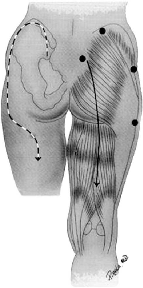
The mainstays of treatment are surgical release and physiologic support, primarily for rhabdomyolysis and secondary renal injury. There are two types of incision that may be used. The classic straight line, shown on the right below, is simple but significantly disfiguring. The question mark incision on the left is kinder and gentler, but more challenging to perform properly.
Bottom line: Compartment syndromes can occur in any enclosed muscle group, which is just about all of them. Always be suspicious if your patient has unexplained elevations of CK, especially if they have tight muscle groups or deep pain in hard to access muscles. Err on the side of checking pressures and releasing those compartments in order to minimize morbidity and ultimate disability.

Reference: Gluteal compartment syndrome: a case report. Cases J. 2:190, 2009.

Lateral Canthotomy For Orbital Compartment Syndrome

I’ve previously written about the orbital compartment syndrome and described the symptoms in the awake patient. I’d like to share a video of the procedure and provide a tip for diagnosing it in unconscious patients.

Patients at risk will have obvious facial trauma. During your physical exam, look for proptosis. This is caused by swelling or hemorrhage behind the globe pushing it forward. It may manifest itself as uneven opening of the eyelids, with the affected side being “propped” open (get it?). But in trauma, there may be significant edema which keeps the lid closed.

The easiest way to observe proptosis is to stand above the patients head, looking toward their feet. Crouch down so you can look across the lids in a direction horizontal to the floor. You should easily be able to detect if one eye protrudes further that the other.

You can also do a poor man’s compartment pressure test by gently using your thumbs to compress both globes simultaneously. If there is substantial difference in resistance between the two sides, a compartment syndrome may be present. Important note! Do a thorough globe exam first! If there is any evidence of globe rupture (hyphema, irregular pupil, extra tissue in the anterior compartment), don’t press the eye or perform a canthotomy. CT scan of the facial bones can help confirm the diagnosis if a mass effect is seen in the orbit or if the optic nerve appears to be on stretch.

The orbital compartment syndrome is an emergency! Once diagnosed, immediately proceed to canthotomy. Otherwise, damage to the optic nerve and retina is likely, and the patient may become blind in the affected eye.

Related post:

Lateral Canthotomy For Orbital Compartment Syndrome

I’ve previously written about the orbital compartment syndrome and described the symptoms in the awake patient. I’d like to share a video of the procedure and provide a tip for diagnosing it in unconscious patients.

Patients at risk will have obvious facial trauma. During your physical exam, look for proptosis. This is caused by swelling or hemorrhage behind the globe pushing it forward. It may manifest itself as uneven opening of the eyelids, with the affected side being “propped” open (get it?). But in trauma, there may be significant edema which keeps the lid closed.

The easiest way to observe proptosis is to stand above the patients head, looking toward their feet. Crouch down so you can look across the lids in a direction horizontal to the floor. You should easily be able to detect if one eye protrudes further that the other.

You can also do a poor man’s compartment pressure test by gently using your thumbs to compress both globes simultaneously. If there is substantial difference in resistance between the two sides, a compartment syndrome may be present. Important note! Do a thorough globe exam first! If there is any evidence of globe rupture (hyphema, irregular pupil, extra tissue in the anterior compartment), don’t press the eye or perform a canthotomy. CT scan of the facial bones can help confirm the diagnosis if a mass effect is seen in the orbit or if the optic nerve appears to be on stretch.

The orbital compartment syndrome is an emergency! Once diagnosed, immediately proceed to canthotomy. Otherwise, damage to the optic nerve and retina is likely, and the patient may become blind in the affected eye.

Related post:

Orbital Compartment Syndrome

Sure, you’ve heard about all the other compartment syndromes: leg, thigh, forearm, buttock, and abdomen to name a few. But how about a compartment syndrome of the orbit?

This isn’t your usual muscular compartment problem, although the basic concept is the same. The eye is surrounded by rigid bones or relatively stiff soft tissue (the eyelid, believe it or not). Any extra tissue or blood added to this compartment dramatically raises the pressure in the area, which is readily conducted to the eye itself. This rapidly results in:

  • Severe eye pain
  • Decreased extraocular movements, which may result in diplopia
  • Proptosis
  • Decreased visual acuity
  • Increased intra-ocular pressure (>40 torr)
  • Slow pupillary response

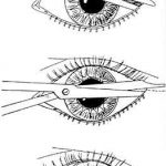
This syndrome should be confirmed rapidly and is one of the few true ophthalmologic emergencies. A lateral canthotomy and cantholysis should be carried out to make the lower lid freely mobile, decompressing the compartment (see diagram). This procedure is not for the faint of heart, but should be familiar in any ED where an ophthalmologist is not readily available. Urgent followup with an eye specialist is mandatory.