All posts by The Trauma Pro

Is The Hybrid OR For Trauma Useful?

Gee, the hybrid OR sounds like a great idea for specific trauma patients. But we’ve seen this before; great idea but doesn’t always translate into a positive result. Is there any literature?

Unfortunately, very little. A group from the University of Calgary in Alberta published a very detailed paper on the nuts and bolts of how they designed their hybrid room from scratch. This paper is very detailed, and the hospital personnel were very thoughtful as they approached the time-consuming and expensive task of designing and building their hybrid room. Of course, they chose a silly acronym as so many do. They called it their RAPTOR room (Resuscitation with Angiography, Percutaneous Treatments, and Operative Resuscitations). Sigh!

Next, they retrospectively analyzed their experience with persistently hypotensive patients arriving at their Level I trauma center over a 17-year period before their hybrid room opened.

Here are the factoids:

  • Of 911 patients, 510 remained persistently hypotensive (SBP<90 torr)
  • 53% (270 patients) were taken directly to OR, usually for laparotomy, thoracotomy, or vascular procedure
  • 29% were admitted to an ICU, 13% to a ward bed, and 5% were taken to interventional radiology (IR)
  • 35 patients (7%) required both OR and IR; the majority had pelvic fractures (77%), the rest had liver lacerations
  • Each case was reviewed, and overall 6% of patients would have clearly benefited from a hybrid room, and 30% would have potentially benefited

Sounds good so far! But we need some more data. Unfortunately, there’s not a lot of it yet. A Japanese group described their experience with treating patients in OR then IR, vs a “hybrid procedure.” This did not involve the use of a true hybrid OR. They moved a C-arm fluoroscopy unit into an OR and part of the procedure was carried out by an interventional radiologist.

And the factoids:

  • A total of 13 “hybrid treatment” patients were compared to 45 who underwent both operation and angiography, but not in the same location
  • Most of the hybrid patients had a laparotomy, but there was a concomitant thoracotomy in one and a craniotomy in another
  • The actual survival in the hybrid patients was 85%, while TRISS predicted that only 62% would live
  • There was no difference in transfusion volumes between the two groups, but total procedure time was significantly shorter in the hybrid group (4 hours vs 6 hours)

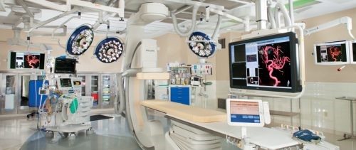
Okay, sounds promising. A second Japanese paper was published last year with much larger numbers. Their hybrid OR was actually a hybrid ER! They installed a multi-slice interventional radiology/CT unit in their resuscitation room! Here are the key findings:

  • A total of 696 patients were reviewed over an 8-year period – 336 hybrid and 360 conventional
  • Mortality was very significantly decreased in the hybrid group
  • OR start was significantly shortened from 68 minutes to 47

Here’s an image of their setup:

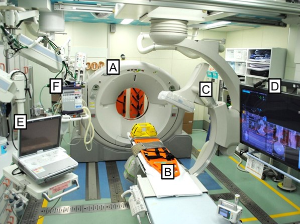
Key: A – mobile CT scanner, B – CT / OR table, C – mobile C-arm, D – 56” monitor, E – ultrasound, F- ventilator

Bottom line: This is quite a unique room. Unfortunately, it is not ideal because it is small and cramped. It looks like it would be difficult to fit more than one surgical team in the room. However, the results look good.

We are finally starting to see objective data involving a reasonable number of patients. A minority of trauma programs have a hybrid OR available to them, and the number of patients who would benefit from it is low. But if a patient needs it, this setup can be life-saving. So who are those patients, exactly?

In my next post, I’ll review a very new (2020) paper specifically on the hybrid room for trauma.

References:

  1. The evolution of a purpose designed hybrid trauma operating room from the trauma service perspective: The RAPTOR (resuscitation with angiography percutaneous treatments and operative resuscitations). Injury 45:1413-1421, 2014.
  2. The potential benefit of a hybrid operating environment among severely injured patients with persistent hemorrhage: How often could we get it right? J Trauma 80(3):457-460, 2016.
  3. Hybrid treatment combining emergency surgery and intraoperative interventional radiology for severe trauma. Injury 47:59-63, 2016.
  4. The Survival Benefit of a Novel Trauma Workflow that Includes Immediate Whole-body Computed Tomography, Surgery, and Interventional Radiology, All in One Trauma Resuscitation Room. Ann Surg 269(2):370-376, 2019.

Why Use A Hybrid OR For Trauma?

Trauma is a surgical disease, and specifically, a disease of bleeding. So many of the tools and processes we have developed for its management revolves around the control of hemorrhage.

When a major trauma patient arrives in the resuscitation room, the initial management involves rapid assessment and correction of life-threatening conditions. Recognition of bleeding is paramount. A rapid decision must be made as to the source of hemorrhage and the best way to control it.

Traditionally, bleeding control has been relegated to the operating room. Body cavities are opened as appropriate, and exsanguination is controlled by clamping, repairing, and/or suturing.

However, some body regions are much more challenging. The most notable is the pelvis, and specifically, the unstable pelvis. In the old days, after wrapping or applying an external fixator, the best we could do was to ligate the internal iliac arteries bilaterally and hope the bleeding would slow down sufficiently (it never really stopped) so that internal packing might have a chance.

As the use of interventional radiography grew in trauma, it became possible to noninvasively occlude the internal iliacs. And then, the radiologists became skilled enough that they could selectively identify and embolize more distal bleeding vessels that would dramatically shut down pelvic bleeding.

But this introduced a conundrum. OR vs IR? Where to go after the trauma bay? I’ve long said that the only place an unstable trauma patient can go is to the OR. Not CT, and certainly not the radiology department.

Only the OR, because that’s the only place that something can actually be done about the bleeding. However, that’s not entirely true now.

Here’s the traditional algorithm for a patient with hemorrhage from pelvic fractures:

They go to the operating room OR interventional radiology. If they start in the operating room and can be stabilized (think external fixation and/or preperitoneal packing), then they might be able to be packaged and taken to IR for embolization. And likewise, if they were initially stable enough to go to IR but crash there, then they must immediately be taken to OR.

But what if you could do both in one room?! That’s the beauty of the hybrid room! It is entirely possible to do two, three, and maybe more cases on the same patient in the same room. Hence, the hybrid OR.

Next post, is the hybrid OR for trauma useful?

What Is A Hybrid OR, Exactly?

A hybrid operating room is a special suite that allows advanced imaging to be carried out at the same time as one or more additional operative procedures. It’s that simple. It contains specialized imaging equipment including fluoroscopy and infusion equipment for radiographic dye administration. Some also contain CT and/or MRI capabilities, although the shielding required for these makes them very rare. It is generally stocked with a variety of endovascular

devices and supplies. The usual anesthesia circuits are available, as are selected surgical packs, typically related to vascular and CV surgery.

These suites are typically large, and can easily accommodate multiple operating teams. However, they are very expensive in a number of ways.

First, they take up a great deal of space. Many have the square footage of two or more standard operating rooms. Initial construction costs are very high, as are remodeling and maintenance costs. They can also tax the hospital engineering infrastructure, from electrical to plumbing to ventilation.

But if a hybrid room is available, it can deliver significant benefits to the hospital and to patient care. Intraoperative imaging can provide immediate quality assurance, and patients can undergo more complex procedures and enjoy a shorter length of stay.

Next post, why use a hybrid room for trauma?

The Hybrid OR, Revisited

Over a year ago I published a series on using the hybrid room for trauma cases. In the meantime, some new papers have been published on this concept. Over the next two weeks, I’ll be refreshing and republishing this information to help you optimize the use of yours, or to support your efforts to get one if you don’t.

While quite a bit of trauma care is routine, involving simpler, single system injuries, a small subset of our patients sustains major, multi-system, and life-threatening ones. They require rapid access to skilled trauma professionals and advanced resources including imaging, operating rooms, and other procedures.

In most trauma centers, initial resuscitation takes place in a trauma resuscitation room in or near the ED. Some diagnostic imaging can be performed there, but more sophisticated studies may require a short (or longer) road trip. Operating rooms and other procedural areas are also usually more distant. And most importantly, each of these areas is designed for a single discipline. Diagnostic radiology has equipment, technicians, and radiologists available. Interventional radiology contains the specialized equipment needed for this more invasive procedure. ORs are designed specifically for surgical procedures, and frequently contain equipment for a single surgical discipline.

But some of our patients require it all! Think about a patient who arrives after a major car crash. Blood pressures are soft, the pelvis is grossly unstable, FAST exam is positive, and there is bleeding from the vagina.

How do we prioritize? Where do we go first? How long will it take the interventional radiology team to arrive? Where’s that external fixator equipment? Can we slip in a CT scan? Where’s OB/GYN??

The solution is right under our nose! Many hospitals have added so-called “hybrid ORs” to their operating suites in order to address the needs of their vascular and cardiovascular surgeons. The next several posts will cover everything you need to know about this important tool for trauma care. I’ll review:

  • What is a hybrid OR, exactly?
  • Why use a hybrid OR for trauma?
  • Is the hybrid OR for trauma useful?
  • Which patients may benefit from a hybrid OR?
  • So you want your own hybrid room?!

Next post: what is a hybrid OR, exactly?

How Quickly Does Hemoglobin Drop After Acute Bleeding?

We all know that hemoglobin / hematocrit drop after blood loss. We can see it decreasing over the days after acute bleeding or a major operative procedure (think orthopedics). And we’ve been told that the hemoglobin value doesn’t drop immediately after acute blood loss.

But is it true? Or is it just dogma?

A reader sent me a request for some hard references to support this. When I read it, I knew I just had to dig into it. This is one of those topics that gets preached as dogma, and I’ve bought into it as well.

Now, I have personally observed both situations. Long ago, I had a patient with a spleen injury who was being monitored in the ICU with frequent vital signs and serial blood draws (but I don’t do that one anymore). He was doing well, then became acutely hypotensive. As he was being whisked off to the OR, his most recent hemoglobin came back at 10, which was little changed from his initial 11.5 and certainly no independent reason to worry.

But hypotension is a hard fail for nonoperative solid organ management. In the OR, anesthesia drew another Hgb at the end of the case, and the value came back 6.

Similarly, we’ve all taken care of patients who have had their pelvis fixed and watched their Hgb levels drop for days. Is this anecdotal or is it real? The doctor / nursing / EMS textbooks usually devote about one sentence to it, but there are no supporting references.

I was only able to locate a few older papers on this. The first looked at the effect of removing two units of red cells acutely. Unfortunately, the authors muddied the waters a little. They were only interested in the effect of the lost red cell mass on cardiac function, so they gave the plasma back. This kind of defeats the purpose, but it was possible to see what happened to Hgb levels over time.

Here were there findings over time for a group of 8 healthy men:

Time Hbg level
Before phlebotomy 14.4
1 week after 11.7
4 weeks after 12.6
8 weeks after 13.6
16 weeks after 13.9

So the nadir Hgb value occurred some time during the first week after the draw and took quite some time to build back up from bone marrow activity.

That’s the longer term picture for hemoglobin decrease and return to normal. What about more acutely? For this, I found a paper from a group in Beijing who was trying to measure the impact of Hgb loss from a 400cc blood donation on EEG patterns. Interesting.

But they did do pre- and post-donation hemoglobin values. They found that the average Hgb decreased from 14.0 to 13.5 g/dl during the study, which appeared to be brief. Unfortunately, this was the best I could find and it was not that helpful.

Bottom line: Your patient has lost whole blood. So, in theory, there should be no Hgb concentration difference at all. But our bodies are smart. The kidneys immediately sense the acute hypovolemia and begin retaining water. The causes ongoing hemodilution within seconds to minutes. Additionally, fluid in the interstitial space begins to move into the vascular space to replace the volume lost. And over a longer period of time, if no additional fluid is given the intracellular water will move out to the interstitium and into the vascular space.

But these things take time. There is an accelerating curve of hemodilution that takes place over hours. The slope of that curve depends on how much blood is lost. A typical 500cc blood transfusion will cause a 0.5 gm/dl drop over several minutes to an hour. We don’t have great data on the exact time to nadir, but my clinical observations support a hyperbolic curve that reaches the lowest Hgb level after about 3 days.

Unlike this curve, it levels off and slowly starts to rise after day 3-4 due to bone marrow activity.

The steepness of the curve depends on the magnitude of the blood loss. After a one unit donation, you may see a 0.5 gm/dl drop acutely, and a nadir of 1 gm/dl. In the case of the acutely bleeding patient with the spleen injury, the initial drop was 1.5 gm/dl. But two hours later it had dropped by over 5 gm/dl. 

Unfortunately, the supporting papers are weak because apparently no one was interesting in proving or disproving this. They were more interested in cardiac function or brain waves. But it does happen. 

Here’s the takeaway rule:

In a patient with acute bleeding, the initial hemoglobin drop is just the tip of the iceberg. Assume that this is only a third (or less) of how low it is going to go. If it has fallen outside of the “normal” range, call for blood. You’ll need it!

References:

  1. Effect on cardiovascular function and iron metabolism of the acute removal of 2 units of red cells. Transfusion 34(7):573-577, 1994.
  2. The Impact of a Regular Blood Donation on the Hematology
    and EEG of Healthy Young Male Blood Donors. Brain Topography 25:116-123, 2012.