Category Archives: Complications

Tips For Avoiding Missed Injuries

In the last two posts, I’ve examined the phenomenon of “delayed diagnosis” or missed injury. I believe that there are only two fundamental reasons why this occurs:

  • Insufficient diagnostic technique – A good physical exam and/or specific diagnostic techniques were not performed. Or rarely, the injury cannot be readily detected by existing techniques and technology. The former is usually the real problem and may be an issue with either the physical exam completeness and/or technique, or judgment used to obtain the appropriate diagnostic test. Example 1: a penetrating injury to the back is missed because the patient is not logrolled to examine this area. Example 2: a spine fracture is missed in an elderly patient with a fall from standing because the back pain found on physical exam is evaluated only with conventional imaging of the spine, not CT.
  • Failure to recognize the injury – The injury was actually identified on a test but was not appreciated by the clinician. Example 1: the radiologist may not have appreciated and reported out a subtle anomaly in the cervical spine imaging. Example 2: you fail to check your patient’s lab tests and miss a sudden spike in serum amylase or lipase the day after your patient was kicked in the epigastrium by a horse.

So what can you do to avoid this potential problem? Here are some tips:

  • Admit that it can really happen to you. If the missed injury rate at your center is off the low end of the bell curve (< 5%), then you are either really good or really blind. You’d better take a close look at your performance improvement process because you may be fooling yourself.
  • Adopt a firm definition of “delayed diagnosis.” Basically, you need a time frame after which a new diagnosis is considered “delayed.” It should be a reasonable time interval after the patient has left the ED. If it’s too short an interval (e.g. once they leave the ED), your number will be unnecessarily high. If it’s too long (days and days later), then significant morbidity may occur that you don’t account for. Most centers have adopted 24, 36, or 48 hours after patient arrival.
  • Implement a tertiary survey process. This is a complete physical re-examination followed by a review of all diagnostic studies (lab and radiology) that have been performed. This exam needs to be dated and timed to ensure that it is performed within the time frame noted above. If a new finding is discovered on the tertiary survey, it is not considered a delayed diagnosis. If found after the survey (or after the pre-determined time interval), it is and must be entered into your performance improvement process.
  • Be paranoid. I hate the phrase, “maintain a high index of suspicion” because it’s meaningless. It’s like those stupid “start seeing motorcycles” bumper stickers. You can’t see what you can’t see. But you can be suspicious all the time, constantly looking for the inevitable clinical surprises of trauma care.

Are There Really More Missed Injuries After Hours?

In my last post, I wrote about the usual reasons for delayed diagnosis: insufficient diagnostic technique or insufficient recognition. What about the time of day? An interesting paper looked at the correlation between admission time and the rate of missed injuries.

The work was done at a large teaching hospital and Level I trauma center in Australia. A large number of patients were reviewed over an 11-year period. The study was complicated slightly by implementing a dedicated trauma unit in the middle of the study period, but the authors stratified their groups to account for this.

Results were stratified by time of admission: office hours, after-hours, and weekends. Missed injuries were defined as those found after the completion of the primary and secondary surveys. The overall statistical treatment appeared to be robust.

Here are the factoids:

  • A huge number of patients (53,000) were reviewed. This is a busy place!
  • There were 2519 missed injuries in 1262 patients (2.4%) [low!]
  • Missed injuries occurred during office hour admissions in 2.2%, after-hours in 2.6%, and on weekends 2.5% of the time
  • The increased incidence of delayed diagnosis in after-hours admits was marginally significant (p = 0.048)
  • Missed injuries appeared to have increased over time and were 1.34 times more likely at the end of the study period vs the beginning
  • Thoracic spine and abdominal injuries were most commonly missed

Bottom line: Hmm, time of day was not on my list of reasons for missing diagnoses. What gives? If you read the article closely, the trauma service at this hospital was staffed with a higher number of trainees after hours and on weekends than during office hours. It was also noted that incomplete physical examination was thought to be a factor in many of the delays. Most likely, both of my listed reasons were in play here. Inexperienced clinicians and insufficient examination are both major factors. And what about the increase in missed injuries over time? Midway through the study, the hospital implemented a dedicated trauma unit, and a tertiary exam became routine. This identified more injuries after the primary and secondary surveys were complete. 

In the next post, I’ll review strategies to decrease the incidence of missed injury.

Reference: Office hours vs after-hours: do presentation times affect the rate of missed injuries in trauma patients? Injury 2015, in press.

Missed Injury / Delayed Diagnosis

Missed injuries (or delayed diagnosis in polite conversation) are the bane of any trauma program. Trauma professionals want to know that they’ve identified all significant injuries in their patients so no future harm will occur due to them.

But what exactly is a missed injury? The definitions tend to vary a bit, which is why their incidence varies so widely in the literature (1 – 39%). The simplest way to describe one is any injury that is identified after a set amount of time. But what is a reasonable time frame? Some define it as the time spent in the emergency department (highest incidence). Others count any injury found after a predetermined period of time (typically 24-48 hours). Some use even longer time intervals, so they obviously look the best and have the lowest incidence.

And what are the factors that contribute to us “missing” these injuries? As you can imagine, there are quite a few, but they boil down to two major categories:

  • Inadequate diagnostic technique (physical exam and/or technology) – I can’t see it
  • Inadequate recognition – I didn’t think of it

A good physical exam with the focused use of appropriate imaging is paramount. Sure, you could use a shotgun approach and just scan everything. The problem is that CT scans have limitations, but we tend to forget that. So we believe that if we don’t see anything on scan, it must not exist. Wrong! The physical exam may pick up suspicious findings that tell the clinician that a specialized study is necessary to rule a potential injury out.

The failure to recognize that an injury is present can occur with everyone that “touches” the patient. The EMT or physician may not appreciate a subtle injury. The radiologist may miss a problem on the images they read. The surgeon might even fail to notice another injury separate from the one she is operating for. Obviously, experience plays a large part in this factor. Students will fail to appreciate a potential injury that a senior clinician will detect rapidly.

What to do about it? In my next post, I’ll review a paper that tries to correlate missed injuries with time of admission. And finally, I’ll discuss some strategies to try to help keep it from happening to you.

Direct Oral Anticoagulant (DOAC) Reversal: Part 2

In the previous post, I reviewed some basic information on DOAC reversal. Five years ago, it was costly and questionably effective. So what has happened in the meantime?

In this post, I’ll review a big trial the pharma company was excited about and make a few comments.

ANNEXA-I Study

This study sought to evaluate the hemostatic effect of Andexanet administration in patients taking a Factor Xa inhibitor who suffered an intracranial hemorrhage.

Key points in this study:

  • It was a five-year, multicenter, randomized controlled trial
  • Subjects had to have taken their medication within 15 hours of the event, had an intracranial hemorrhage identified by CT within 12 hours of symptoms, and randomized in the study within two hours after the scan
  • There were 263 patients reversed with Andexanet and 267 with “usual care,” which was not clearly defined aside from administration of prothrombin complex concentrate (PCC)
  • Traumatic ICH was only present in about 13% of subjects, and the average volume was about 10 mL. Most were intracerebral hemorrhages (90%), with 5% or less being subdural hematomas.
  • Andexanet treatment was associated with increased “hemostatic efficacy,” a combination variable consisting of volume change, change in NIH Stroke Scale score, and no need for rescue therapy within 12 hours.   There was also decreased hematoma volume change by 3.8mm (12%), an increased number of thrombotic events (10% vs. 6%), and an increased number of ischemic strokes (6.5% vs. 1.5%) at 30 days. There was no difference in deaths at 30 days.
  • Hemostatic efficacy was highest in intracerebral hemorrhages and nearly ineffective for subdural hematomas
  • Hemostatic efficacy was significantly higher than that of patients who received PCC in the “usual care” arm, but it was no better than usual care without PCC (?)

Bottom line: Wow! That’s a lot of numbers. The company was excited because the trial was stopped early due to “superior [hemostatic] efficacy vs usual care.” Basically, what they are saying is that the combination of hematoma size, stroke scale, and lack of need for other rescue therapy was significantly lower in patients treated with andexanet alfa. 

But is this meaningful in trauma? There are several issues, IMHO:

  • The study was not powered to detect mortality or functional outcome differences, which is what we trauma people are really interested in
  • The primary outcome (hemostatic efficacy) was powered mainly by hematoma size change, which is not of any clear clinical significance
  • There were some shenanigans from company involvement in the study design, with several protocol amendments that occurred
  • It was not clear what “usual care” consisted of other than PCC administration in some patients
  • There was no information on costs

In my next post, I’ll cite several systemic reviews and meta-analyses to come to some final conclusions about this drug.

Reference: Andexanet for factor xa Inhibitor–Associated acute intracerebral hemorrhage. N Engl J Med. 2024;390(19):1745-1755.

What Is: A Morel-Lavallee Lesion?

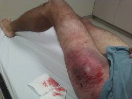
Anyone who takes care of blunt trauma has seen the Morel-Lavallee lesion (M-L). Here’s an obvious one because it’s acute:

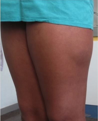
The M-L lesion is essentially a closed degloving injury in which the skin remains intact. The subcutaneous tissue is sheared off of the underlying fascia, and typically blood accumulates in the potential space that is created. This picture shows a less acute lesion; the bruising and ecchymosis on the surface have resolved. Note the collection on the lateral thigh:

These injuries may take a very long time to resolve and may leave some residual deformity. The definitive management has never been very clear: needle drainage vs incision, timing, compression wraps, etc.

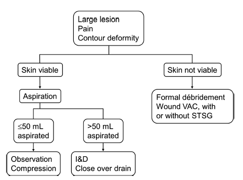
The Mayo Clinic reviewed their 8-year experience with 87 of these lesions to try to shed some light on proper management. They treated their patients in four different ways: needle drainage, incision and drainage, compression wraps, and debridement with vacuum drainage devices. Here are the factoids from their study:

  • Motor vehicle crash was the most common etiology for this lesion, which makes sense due to the energy needed to shear the tissues
  • The most common locations were thigh, hip, and flank
  • The incidence of pre-existing conditions that might influence outcome (diabetes, obesity, smoking history, use of anticoagulants) did not seem to influence outcomes
  • Lesion location did not change the recurrence rate (even over joints)
  • Aspiration suffered the highest recurrence rate (56%) vs only 15-19% in the other groups
  • Aspiration of more than 50cc of fluid was more common in lesions that recurred (83%) vs those that did not (33%)

Their experience led them to develop the following practice guideline:

An incision and drainage procedure is not necessarily straightforward. Many of these wounds develop a pseudo-capsule if they are long-standing. Closure of the dead space can be challenging and may require quilting sutures or use of fibrin glue in addition to low suction drains. Some surgeons use sclerosing agents, either alone or in addition to the adjuncts listed above.

Bottom line: The Morel-Lavallee lesion can be challenging to treat. Although this study has limited numbers, it provides enough guidance to suggest a consistent way of managing it. I recommend adopting this algorithm to provide a standard pathway for dealing with it.

Reference: The Mayo Clinic experience with Morel-Lavallee lesions: establishment of a practice management guideline. J Trauma 76(2):493-497, 2014.