You’ve just received a young male who had been stabbed under his right arm in your emergency department. He’s awake, talking, and very friendly. He met your trauma activation criteria, so you are cruising through the full evaluation. Lines in, blood drawn, clothes off. He wonders aloud if all this is really necessary.
Then, on FAST exam, you see it. A pericardial stripe that looks like a mix of liquid and clotted blood. Your colleague steps in and verifies the exam. But vital signs are normal, the patient is fine.
What next? CT of the chest to further define this? A formal echo to confirm? Your surgeon says no, we’re going to the OR, now! Reluctantly, you package the patient and send him on his way. In the OR, the anesthesiologist takes his time, putting in an arterial line, asking the patient unrelated questions. A thoracotomy? Really? The patient remains awake and alert through all of this.
So here’s the fourth law of trauma:
Even awake, alert, and stable patients die. And it hurts that much more when they do.
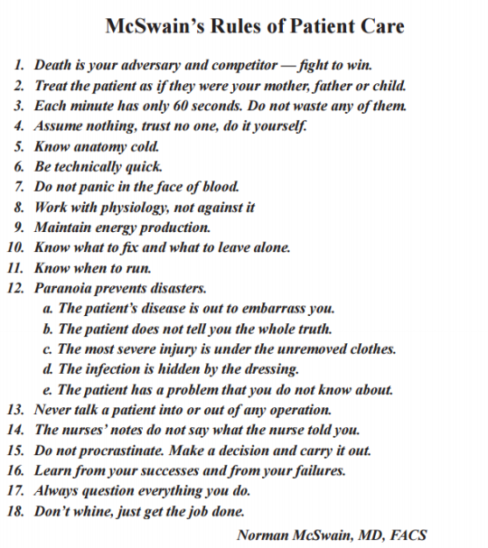
Bottom line: You know the diagnosis in this case. And you know what needs to be done. But the awake and alert patient fools us. Fakes us out. Somehow, we equate the ability to talk intelligently with being fine. But evil things can be going on inside that don’t rear their ugly head until it’s too late. Don’t get suckered! Believe your exam, not what the patient thinks they are telling you.