Tag Archives: rib fractures

WTF? A Fractured Rudimentary Rib?

Is it real, or just another one of those crazy things that radiologists like to add to their reports? I recently came across one of these for the first time in over 30 years of practice. What is it? And is it significant in your management of a trauma patient?

A rudimentary rib is simply an extra one (supernumerary). They can be found on vertebrae where ribs are not supposed to be present, typically C7 and L1. The most common supernumerary ribs are found at C7, and are a well documented cause of thoracic outlet syndrome. 

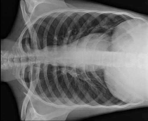
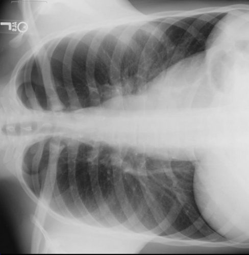
Rudimentary ribs are less commonly found on lumbar vertebrae, and they tend to be longer than the transverse processes. This means that it is possible to break them given moderate to high energy blunt torso trauma. The image below shows a person with 2 rudimentary lumbar ribs on L1.

These are very rare congenital variants. It is more likely that your patient is showing abnormal bone formation after a previous fracture, so question them closely for a history of trauma.

What’s the clinical significance? There’s little chance of hemothorax or pneumothorax. But they cause pain like any other fracture. Just apply your usual routine for rib fracture management: analgesia and pulmonary toilet. Since it takes a relatively large amount of energy to break these short little ribs, be on the lookout for other occult injuries as well.

Bottom line: This isn’t just a weird radiology “red herring.” Rudimentary rib fractures can occur, although a history of previous injury should be ruled out. Manage like any other rib fracture, but beware of potential occult injuries.

Related posts:

Answer: Finding Rib Fractures On Chest Xray

There was a lot of chatter regarding my practical tip yesterday, rotating the chest xray to better visualize rib fractures. Here’s the quiz xray from yesterday: 

And here’s the lateral view:

The fracture is perfectly placed on the most lateral aspect of the left 9th rib. You can download the full size rotated jpg here if you are having a hard time seeing it on the reduced size image above. Piece of cake!

Related post:

Practical Tip: Finding Rib Fractures On Chest Xray

Here’s a neat trick for finding hard to see rib fractures on standard chest x-rays.

First, this is not for use with CT scans. Although chest CT is the “gold standard” for finding every possible rib fracture present, it should never be used for this. Rib fractures are generally diagnosed clinically, and they are managed clinically. There is little difference in the management principles of 1 vs 7 rib fractures. Pain management and pulmonary toilet are the mainstays, and having an exact count doesn’t matter. That’s why we don’t get rib detail x-rays any more. We really don’t care. Would you deny these treatments in someone with focal chest wall pain and tenderness with no fractures seen on imaging studies? No. It’s still a fracture, even if you can’t see it.

So most rib fractures are identified using plain old chest xray. Sometimes they are obvious, as in the image of a flail chest below.


But sometimes, there are only a few and they are hard to distinguish, especially if the are located laterally. Have a look at this image:


There are rib fractures on the left side side on the posterolateral aspects of the 4th and 5th ribs. Unfortunately, these can get lost with all the other ribs, scapula, lung markings, etc.

Here’s the trick. Our eyes follow arches (think McDonald’s) better than all these crazy lines and curves on the standard chest x-ray. So tip the x-ray on its side and make those curves into nice arches, then let your eyes follow them naturally:


Much more obvious! In the old days, we could just manually flip the film to either side. Now you have to use the rotate buttons to properly position the digital image.

Final exam: click here to view a large digital image of a nearly normal chest xray. There is one subtle rib fracture. See if you can pick it out with this trick. You’ll have to save it so you can manipulate it with your own jpg viewer. If you find it, tweet it out to me! Let’s see who gets it first!

Related posts:

CT Scans And Rib Fractures

Last week I discussed the importance of treating rib fractures in older patients with the greatest respect. One reader commented:

“number of rib fratures are not that accurate by x-ray. If further evaluate by CT, more fractures will be identified”

Well, I agree and I disagree. Chest xray is notoriously inaccurate when it comes to diagnosing or counting rib fractures. Some older studies have shown that a plain chest xray may miss as many as 50% of all rib fractures. On the other hand, CT scan is very accurate at diagnosing them.

But the question is, do we need to know exactly how many ribs are fractured? In general, the answer is no. Rib fracture is a clinical diagnosis. A patient with an appropriate mechanism and focal tenderness on the chest wall has a rib fracture unless proven otherwise. Do we need to prove otherwise? No. They still have pain, and it still needs to be treated. The degree of pain and pulmonary impairment determines the need for admission and more advanced therapies, not an exact count of ribs fractured. 

Bottom line: Rib fracture is a clinical diagnosis! CT scan of the chest for diagnosing rib fractures (or pneumothorax, or hemothorax for that matter) is basically not indicated. It delivers a lot of radiation (and IV contrast if you mistakenly order it), but does not change management. For blunt trauma, CT of the chest should only be used for screening for aortic injury. The only possible indication I can think of is to plan ORIF of complicated, displaced rib fractures. But in that case, let your surgical specialist decide if the test is really necessary.

Related posts:

More On Rib Fractures In The Elderly

In response to my post yesterday, Chris Nickson wrote:

“Is it possible there were physiologically young but chronologically old patients with isolated rib #s that were sent home from ED that were not included in the study?

I suspect that there are patients over 65 years old with isolated rib #s that can be safely discharged if follow up is bullet proof and pain well controlled.

However, I agree with your over riding message to not underestimate the elderly rib fracture!”

Very few authors do anything but stratify the elderly by age when they write research papers. They do not look at frailness, even though there are scoring systems to do just that. Plus, the retrospective nature of most of the literature (including this paper) preclude the use of such a scale.

Most of the elderly patients that we all see in the ED are selected out to be frail. The healthy ones stay at home and tough out a single rib fracture or even two. But the ones who are brought in are most likely having issues with pain or breathing, thus prompting the visit.

Bottom line: I agree that some elderly patients (the younger and healthier ones) could potentially be sent home from the ED with some pain medication. But the trauma professional needs to make sure that they are comfortable and can move about with well controlled discomfort. They also need good discharge instructions regarding returning to their primary physician or ED promptly if they start to have pain control or respiratory problems. If there is any doubt, bring them in to the hospital for a brief visit for pain control and pulmonary management.

Related post: